Case Presentation: 59 year old African American male with a history of sarcoidosis on prednisone, pulmonary HTN, CKD 3, NIDDM, and NICM presented to the ED with a 2 week history of leg swelling/pain. He was febrile on presentation. He was prescribed cephalexin for cellulitis and discharged.
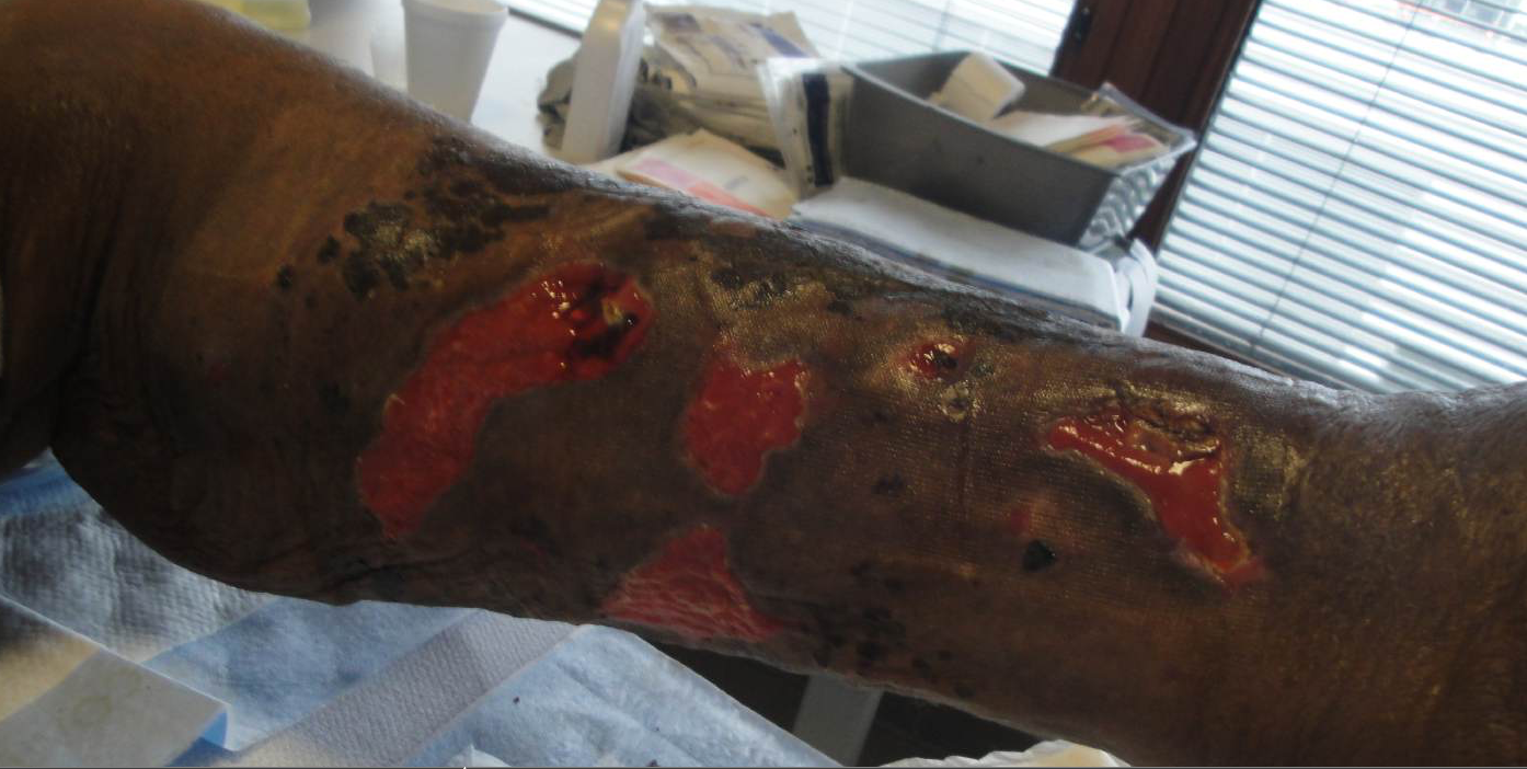
He returned 2 days later with worsening lower extremity edema. Vitals were notable for Temp 100 degrees Fahrenheit and tachycardia (110s). On exam, he had bilateral pitting edema to the mid-thigh. Bilateral lower extremities were warm, erythematous, and tender with multiple, irregular shallow ulcers that had a yellow fibrinous base. The surrounding skin was dusky/black with ill-defined 1-3 cm round dusky patches and indurated subcutaneous nodules. Verrucous dark-brown plaques and deeper calcaneal ulcers were seen on both feet. He was neurovascularly intact. He also had diffuse crackles bilaterally. Labs were notable for leukocytosis (14K), BUN 70 mg/dl, SCr 2.7mg/dl (baseline 1.1-1.3 mg/dL), calcium 9.0 mg/dl, PTH 99 pg/mL, and Vitamin D 25OH 20 ng/mL and CXR showed vascular congestion. Infectious workup and vasculitis serologies were negative. Punch biopsy was remarkable for calcium vessel deposition with minimal vessel occlusion on von Kossa stain. Occlusive fibrin clots were also noted. These findings were consistent with small vessel vasculopathy. Diagnosis of nonuremic calciphylaxis (NUC) was made based on multiple potential triggers (chronic long term steroid use, sarcoidosis) as well as response to sodium thiosulfate. Patient has since had frequent soft tissue infections leading to multiple hospitalizations.
Discussion: Calciphylaxis (calcific uremic arteriolopathy [CUA]) is an infrequent, well-documented phenomenon post-renal transplant and in end stage renal disease. Renal failure was originally believed fundamental to developing this condition. Here we presented a case of presumed nonuremic calciphylaxis likely secondary to sarcoidosis and steroid use. Although biopsy results were equivocal, von Kossa stain was not performed with the addition of Alizarin Red, potentially leading to a decreased sensitivity. However, the history/exam, risk factors and treatment response were suggestive of this disease. Less than 100 cases of NUC are documented, and it is most frequently seen in adult Caucasian women. Pathophysiology remains poorly understood, although histologically it matches CUA. Most cases of NUC are associated with primary hyperparathyroidism, connective tissue disease, alcoholic liver disease, malignancies, and the use of corticosteroids and warfarin. Treatment of choice is sodium thiosulfate, but little data exists to guide duration of therapy. Mortality exceeds 50% secondary to sepsis from superinfection of non-healing ulcers.
Conclusions: NUC is a very rare and devastating condition. As it is so poorly understood, management is based on expert opinion and CUA strategies. In this case, timely diagnosis led to rapid initiation of treatment with sodium thiosulfate.
.png)