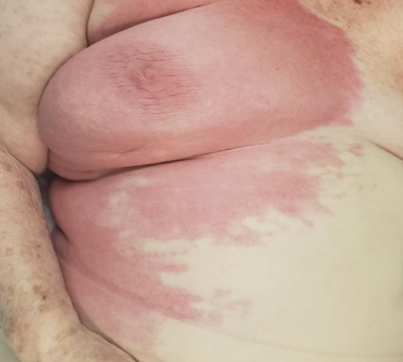
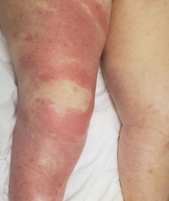
Case Presentation: A 90 year old female with invasive ductal carcinoma of the breast previously treated with lumpectomy and whole breast radiotherapy, currently on hormonal therapy presented to the hospital with 3 days of confusion, slurred speech, generalized weakness and erythema of the right chest and right lower extremity. Patient denied recent changes in skin products, detergents or medications. She had no fever, chills, nausea, vomiting, diarrhea, cough or shortness of breath. Physical exam was notable for a well-demarcated, blanching erythematous rash involving the right breast, axillary area and right lower extremity but sparing the abdomen, the right arm and the left side of the body. The right thigh and leg were warm and tender. The white blood cell count (WBC) was 29.1 K/UL with 78% neutrophils. Cefazolin was initiated and patient underwent a skin biopsy of the rash on the right chest wall. After initiation of antibiotics her rash significantly improved except at the level of the right chest and her WBC plateaued around 18 K/UL. Skin biopsy revealed a dense neutrophilic infiltrate without evidence of leukocytoclastic vasculitis compatible with Sweet syndrome. Oral prednisone at 80mg per day was initiated, cefazolin was transitioned to oral cephalexin and patient was discharged with outpatient follow up with Dermatology.
Discussion: Sweet syndrome, also known as acute febrile neutrophilic dermatosis, is a rare inflammatory disorder characterized by the sudden onset of painful, edematous and erythematous plaques. Sweet syndrome mimics cellulitis with fever, leukocytosis and neutrophilia being the main clinical manifestations in addition to the skin lesions. Skin findings are typically asymmetrical in distribution, with the upper extremities, trunk, head, and neck being the typical sites of involvement. Lower extremities are less often involved. Lesions are usually several millimeters to centimeters in diameter, but large cellulitis-like plaques have been reported as a rare clinical presentation of Sweet syndrome, as seen in our patient. Sweet syndrome is classified into classical (also called idiopathic), malignancy-associated and drug-induced Sweet syndrome. Our case is consistent with classical Sweet syndrome, as the evaluation for malignancy was negative and patient was not on any medications associated with this disorder including G-CSF. Its pathogenesis remains uncertain but it’s thought to be secondary to immune reaction to different antigens with concomitant cytokines or chemokines dysregulation. Skin biopsy is required to confirm the diagnosis by detecting histopathologic evidence of a dense neutrophilic infiltrate without leukocytoclastic vasculitis. Systemic corticosteroids is the mainstay therapy. Prednisone is recommended at 0.5 to 1mg/kg per day initially, followed by a tapering dosage to prevent recurrence. Clinical improvement is expected after a few doses and complete resolution at 1-2 weeks of treatment. When secondarily infected, Sweet syndrome lesions can partially improve if treated with antibiotics directed against staphylococcus species.
Conclusions: Clinicians should have a high index of suspicion for Sweet syndrome when assessing a patient with fever, neutrophilic leukocytosis and erythematous skin plaques with features not typical of cellulitis such as distant lesions, asymmetrical distribution, truncal involvement and lack of response to standard antibiotics. If Sweet syndrome is suspected, a dermatology consult and skin biopsy should be considered.