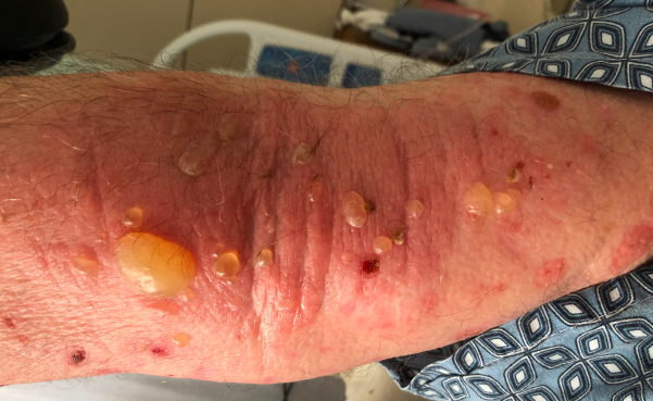
Case Presentation: A 76-year-old man with history of diabetes mellitus, atrial fibrillation, colonic adenoma, and recent self-reported bed bug infestation presents with one month of pruritic rash. On interview, he also endorses progressive edema, dyspnea on exertion, orthopnea, as well as 5 days of melena. Admission exam is notable for anasarca and diffuse, tense fluid-filled blisters overlying erythema on proximal legs, torso, and distal arms. Labs are notable for a leukocytosis of 13.8 K/uL with 20% eosinophilia (absolute eosinophilia of 3260 cells/uL), hemoglobin of 8.7 g/dL (from 15.6 g/dL one year prior), and pro-BNP 1212 pg/mL. Dermatology is consulted for his rash, which is determined to be bullous pemphigoid based on biopsy as well as biomarkers (BP 180 Antibody positive). CT angiogram of the Chest/Abdomen/Pelvis reveals diffuse adenopathy, two hypodense liver lesions, mild pulmonary edema, and mild anasarca. EGD demonstrates ulceration at the GE junction. Liver biopsy demonstrates poorly differentiated adenocarcinoma. Ultimately, his clinical picture is felt to be secondary to metastatic gastroesophageal junction cancer with likely paraneoplastic bullous pemphigoid. He is started on high potency topical steroids, which significantly improves his bullous pemphigoid rash by discharge.
Discussion: Bullous pemphigoid is an uncommon autoimmune subepithelial blistering disease that is estimated to occur in 4-22 cases per million individuals per year. Bullous pemphigoid primarily occurs in patients over 60 years old as well as those with two or more comorbidities. Additionally, it has associations with medications, neurological disorders (dementia, Parkinson’s disease, bipolar disorder), as well as malignancy (particularly hematologic). The pathogenesis is not fully understood but is proposed to involve autoantibody-mediated damage to the epithelial basement membrane zone with resultant separation of the epidermis from the dermis. This forms the characteristic 1-3 cm tense bullae on an erythematous, urticarial, or noninflammatory base with severe pruritus that are associated with bullous pemphigoid. Diagnosis is primary based off of skin biopsy with direct immunofluorescence demonstrating linear IgG and/or linear C3 staining along the basement membrane zone, which is present in more than 90% of cases. However, clinicians can also check bullous pemphigoid antigens 180 and 230 (two principal basement membrane zone antigens). Initial treatment includes the use of high-potency topical steroids. However, if topical steroids are not feasible, then treatment with systemic glucocorticoids or other immunosuppressants can be utilized.
Conclusions: In Hospital Medicine, rashes are frequently encountered on the wards. However, one study found that the primary ward team only determined an accurate dermatologic diagnosis in less than 25% of cases. As such, familiarity with life-threatening or severe rashes is important – particularly if the diagnoses can help lead to an underlying pathology. Bullous pemphigoid is representative of one of these rashes that are important to recognize as a hospitalist. Bullous pemphigoid is an uncommon autoimmune disease leading to tense bullae through damage of the epithelial basement membrane. This patient demonstrates the association between malignancy and bullous pemphigoid and serves as an important dermatologic case for hospital medicine clinicians.