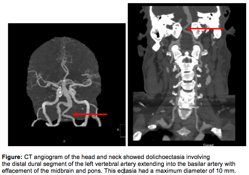
Case Presentation: A 58-year-old man presented with acute onset of chest pain, vertigo, and ataxia, described as “falling to the right as objects moved to the left.” History included coronary artery disease requiring multiple stents and remote cerebrovascular accident with residual left upper extremity weakness. Exam was notable for vertical nystagmus. EKG showed left atrial enlargement and left anterior fascicular block, unchanged from prior studies. CT angiogram of the head and neck (Figure) showed dolichoectasia to a diameter of 10mm involving the distal dural segment of the left vertebral artery, extending into the basilar artery with effacement of the midbrain and pons. MRI brain with contrast showed no evidence of intracranial abscess, acute infarct, or hemorrhage. No surgical intervention was indicated, and he was discharged with recommendations for blood pressure and lipid control.
Discussion: Vertebrobasilar dolichoectasia is an anatomic abnormality characterized by expansion and increased tortuosity of the vertebrobasilar arteries. Prevalence is believed to be less than 0.05% of the population worldwide. The condition is usually discovered by imaging after patients develop ischemic stroke, brainstem compression, or hydrocephalus. This is a rare case where this vascular anomaly was discovered from an interesting presenting constellation of ataxic symptoms.The scarce literature for symptomatic dolichoectasia points control of arterial hypertension as the best strategy to prevent enlargement of ectasia and decrease the incidence of ischemic stroke and intracranial hemorrhage. It remains unclear if the patient in the case would have benefitted from surgical intervention as he improved from medical management and physical therapy.
Conclusions: Intracranial dolichoectasia can masquerade as vertigo with an otherwise unremarkable presentation. Very few patients go through microsurgical treatment, which entails removal of expanded arteries and replacement with new blood vessels. Hope remains for advancement of endovascular and neurosurgical techniques to deal with enlarging dolichoectasia.