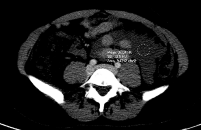
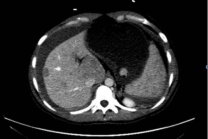
Case Presentation: A 31 year-old woman with a history of portal vein thrombosis at age 8 with cavernous transformation resulting in non-cirrhotic portal hypertension presented with acute onset of upper abdominal pain. The patient was pale, tachycardic, with diffuse abdominal distention and tenderness. Laboratory data was significant for an acute drop in hemoglobin from 14 to 7.4 g/dL. CT scan revealed multiple low attenuating lesions in the liver concerning for liver metastatic disease and heterogenous appearance of ascites which had mixed attenuation though no layering seen with hemorrhage, most concerning for omental thickening or peritoneal carcinomatosis. CT chest did not show any pulmonary lesions and AFP, beta HCG, CA 19-9, and CEA were all within normal limits. A diagnostic paracentesis revealed frank blood with 2.9 million RBCs/L. CT angiogram showed no active bleeding but was concerning for a rapidly expanding liver lesion that ruptured. IR particle embolization of a left hepatic liver lesion was performed. After monitoring for several days to ensure stability, the patient underwent a 5.3L paracentesis and liver biopsy. Pathology revealed a diagnosis of multifocal hepatic adenomas, also called hepatic adenomatosis.
Discussion: Hospitalists frequently encounter new liver lesions or masses and formulate a working differential. The clinical scenario above emphasizes on the following learning points. The first teaching point is that we’re taught to think about rapidly growing lesions as cancerous; however it is important to include other causes such as focal nodular hyperplasia, hepatic adenomas, or hemangiomas. The rupture and ultimate hemoperitoneum caused by her lesion was very informative. Hemangiomas and focal nodular hyperplasia have a very minimal risk of rupture. Patients with hepatocellular carcinoma have a 3-15% risk of rupture and typically take minimally a month to double in size. This information brought hepatic adenomas higher up on our differential. About 25 – 64% of adenomas will bleed.Another teaching point is that risk factors may not be helpful in weighing differentials. Our patient was not obese nor had hepatic steatosis or metabolic syndrome – all risk factors for hepatic adenomatosis. She did not have the other risk factors for adenoma development such as use of estrogen, anabolic androgens, genetic syndromes such a glycogen storage disease or familial adenomatous polyposis.The last teaching moment is the double edged sword of a cancer diagnosis as a differential. It was quickly thought the patient had possible hepatocellular carcinoma with peritoneal carcinomatosis or an unknown primary cancer with metastatic disease to the liver. This led to her placement on an oncology service and an extensive work up which included a CT chest, pelvic ultrasound, tumor markers, and an oncology consult. If any clinician encountered a similar case as above, with knowledge of our patient’s result, it would still be best practice to obtain an extensive work up to not miss a detrimental diagnosis such as cancer. However, interpreting the behavior of the lesions and advocating to obtain tissue as quickly as possible may be beneficial. Our patient was transferred to a hepatology service and discharged with instructions to have surveillance imaging every 3 months to monitor for malignant transformation and potential need for a liver transplant.
Conclusions: Specific characteristics of masses are useful to differentiate between malignant and benign etiologies – such as the growth or rupture of liver lesions.