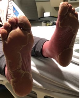
Case Presentation: A 55yo woman with no prior history of rheumatologic disease or skin disorders presented to the hospital with erythroderma. Two months prior to presentation, she had a sudden eruption of patchy, erythematous plaques and diffuse scaling of the scalp, face, upper chest, and back. This progressed to involve her entire torso and extremities. Notably, approximately 3 weeks prior to the onset of the rash, she was diagnosed with mildly symptomatic COVID-19 infection. She was seen in the outpatient dermatology clinic and laboratory examination revealed positive ANA of 1:320, negative DsDNA, ENA, and hepatitis viral panel. Two skin biopsies were performed, she was diagnosed with cutaneous lupus and started on oral prednisone, hydroxychloroquine, and mycophenolate. On physical examination, she had diffuse erythema encompassing approximately 90% body surface area, erythematous plaques with islands of sparing, and waxy keratoderma of the palms and soles. Dermatology was consulted and performed a skin biopsy of an abdominal lesion. This revealed psoriasiform dermatitis with alternating orthokeratosis and parakertasosis and follicular plugging, consistent with a diagnosis of pityriasis rubra pilaris. She was discharged from the hospital on acitretin, topical triamcinolone, and prednisone was tapered off. She was seen in clinic one month after discharge without improvement in symptoms and was started on ixekizumab.
Discussion: Pityriasis rubra pilaris (PRP) is a rare inflammatory dermatosis of unknown etiology, but has been associated with malignancy, autoimmune disorders, and infections and in fact may be the primary manifestation of HIV infection (2, 4). Because PRP is unresponsive to systemic steroids and has debilitating sequela that frequently results in poor quality of life and depression (1), early recognition and diagnosis is important. PRP is classified into six subtypes, based on Griffith’s classification, with variable presentations and natural history. Though it may be confused with other erythrodermic disorders, especially psoriasis, PRP has characteristic physical and histologic findings that aid in diagnosis. In adults, PRP usually starts on the face and scalp with a cephalocaudal progression and skin lesions have interspersed areas of uninvolved skin known as “islands of sparing (3).” Nail changes occur, but unlike psoriasis, onycholysis, and nail pitting are usually absent (2). Typical histopathologic findings include alternating horizontal and vertical parakeratosis and hyperorthokeratosis, keratotic plugging, and a predominantly lymphocytic dermal infiltration (2,3). Though response to treatment varies depending on the subtype, response is better achieved with combining topical (emollients, steroids, retinoids) and systemic (retinoids, methotrexate, TNF-alpha inhibitors) therapies (3).
Conclusions: While hospitalists might be familiar with various post-infectious, drug-induced, and auto-immune cutaneous manifestations, PRP is a rare culprit that is usually not considered in patients presenting with rash after COVID-19 infection. Given that it is frequently misdiagnosed as systemic cutaneous lupus or psoriasis (3, 5), knowledge of its characteristic physical examination and histopathologic features is important.