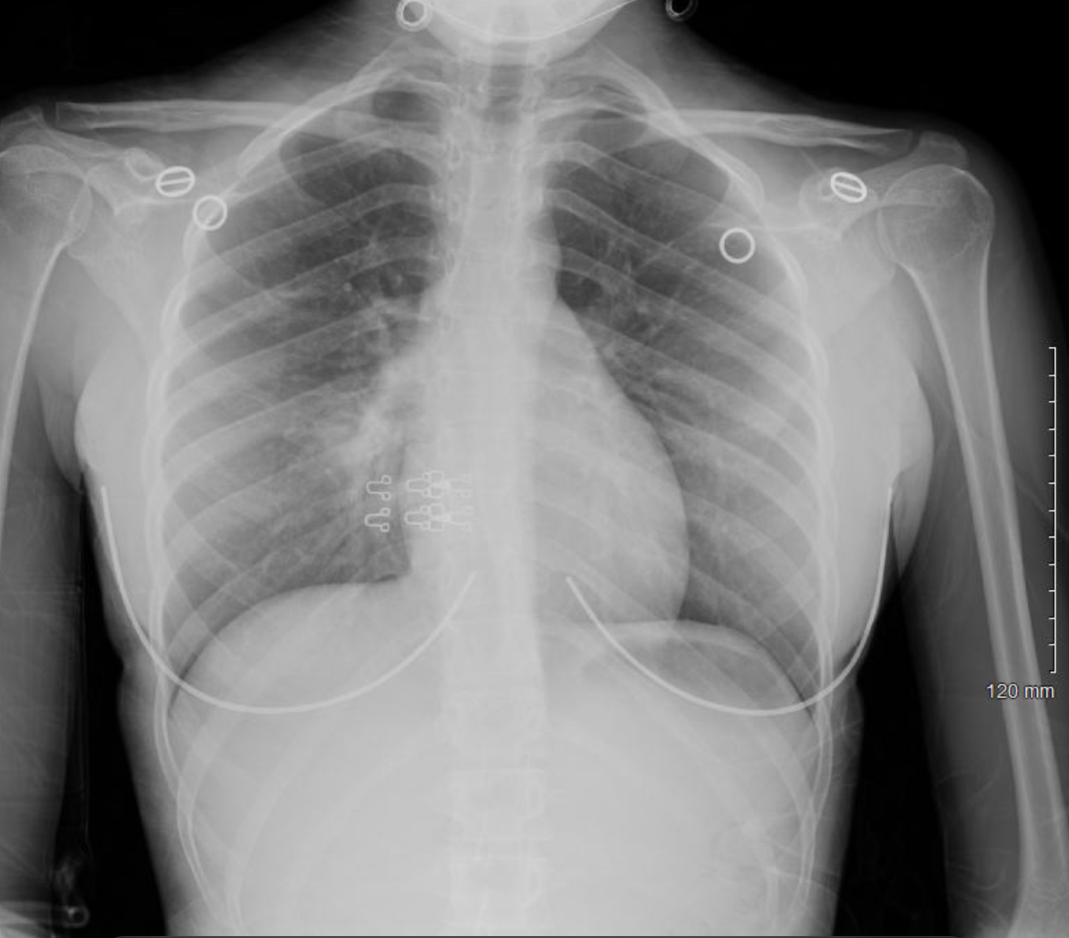
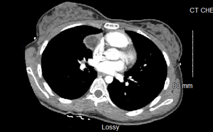
Case Presentation: A 17-year-old female with a past medical history of allergic rhinitis presents as transfer from an outside hospital for hemoptysis and chest pain. She reports three episodes of ¼ cup worth of bright red blood occurring within the last six months. No reported triggers and no history of trauma, smoking, vaping, lung or HEENT disease. She also relays a three-pound unintentional weight loss. Chest pain was located in the middle and right-side of chest and was described as feeling “hard to breath or expand my chest.” Chest pain occurred only with episodes of hemoptysis and was associated with anxiety and palpitations. Three months prior, she presented with similar symptoms to an outside ED where she was diagnosed with pneumonia on imaging and treated with a 3-day course of azithromycin. Physical examination was significant for a thin-appearing adolescent with a BMI in the 4th percentile. She was afebrile, hemodynamically appropriate for age, and saturating well on room air without any respiratory distress. No obvious sources of bleeding were present on exam. Chest radiograph showed a subtle residual ground-glass density noted in the right infrahilar lung. A d-dimer, EKG, and TB test were negative. CT chest with contrast demonstrated a complex right pericardial cyst measuring 3.0 x 2.4 x 6.0 cm extending into the right middle lobe. Trans-thoracic echocardiogram demonstrated an extra-cardiac mass lateral to the right atrium that could be seen moving with the atrium. Serum hCG, AFP, and LDH were negative. Pediatric surgery and cardiothoracic surgery were consulted for surgical resection of the mass with gross pathology consistent with dermoid cyst. Final surgical pathology consistent with 5.7 cm mature cystic teratoma with teratoma-bronchiolar fistula.
Discussion: This adolescent patient presented with hemoptysis, chest pain, and an opacity of the right middle lobe on chest radiograph. With initial evaluation, symptoms were suspected to be due to infectious etiology and were treated with empiric antibiotics. Prior evaluation had also contributed anxiety as the cause of her chest pain in the setting of a normal EKG and negative d-dimer. Re-evaluation and further imaging characterized it as a mass in the anterior mediastinum. Surgical resection revealed a mature cystic teratoma and was subsequently curative. Hemoptysis was likely secondary to post-obstructive pneumonitis from the lesion. While a teratoma of the mediastinum is considered a rare germ cell tumor, the anterior mediastinum is the most common extra-gonadal site. While it is slow growing and benign, it can cause symptoms of cough, dyspnea, and chest pain as a result of compression of nearby structures. Tumor rupture can result in hemoptysis, respiratory distress, or cardiac tamponade.
Conclusions: Hemoptysis in the pediatric population holds a wide differential, including infectious, cardiac disease, pulmonary vascular disorders, bronchiectasis, trauma, tumors, medications, and toxins. While respiratory infections are the most common cause, it is important to consider all causes. Prompt emergent evaluation is needed in unstable patients. In the adolescent population, it is important to consider toxin exposure, such as vaping, electronic cigarettes, and illicit drug use. In chronic and recurrent symptoms, further evaluation with more detailed imaging or referral is likely warranted. This patient case is an example of the importance of a wide and unbiased differential in the adolescent population.