Background: Left atrial enlargement (LAE) is a common disorder that is associated with cardiovascular morbidity and mortality. Cardiac point-of-care ultrasound (POCUS) is frequently used to assess for LAE. However, prior studies reported only modest test characteristics with sensitivity, specificity, and 60-75% accuracy using outdated gold standard reference ranges. This study was designed to determine the test characteristics of LAE assessment in the parasternal long axis (PLAX) view by non-cardiologists POCUS users in comparison to the gold standard of left atrial volumetric index (LAVI) as reported by transthoracic echocardiography (TTE.)
Methods: This study included consecutive adult, complete TTEs performed at the University Medical Center. Exclusion criteria included duplicate exams from the same patient, studies with missing reported LAVI data, and studies with reported altered cardiac anatomy (heart transplant, mechanical valve replacements, left ventricular assist devices, and intra-atrial devices.) The power analysis estimated a sample size of 240 TTEs. Selected exams were de-identified and exported for review by 3 blinded Hospital Medicine faculty physicians with POCUS expertise. Demographic information (age, sex, height, and weight) and the reported cardiac findings on the TTE report (LAVI, chamber measurements) were recorded. Images in the PLAX view were assessed using two methods. Using the first method, reviewers visually estimated the anteroposterior diameter of the left atrium and compared it to the overlying ascending aorta throughout the cardiac cycle. The second approach involved a semi-quantitative measurement of the LA anteroposterior diameter in end-ventricular systole with a 4 cm cut-off defining LAE. IRB approval was obtained before the study was initiated.
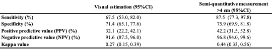
Results: Three hundred twenty-one TTE studies were initially selected, and 241 (75%) met the inclusion criteria. A total of 14.9% of the studies had moderate to severe LAE. The visual estimation method had a sensitivity of 67.5% [95%CI: 53.0%, 82.0%] and a specificity of 71.4% [95%CI: 65.1%, 77.6%] (Table 1.) The semi-quantitative approach had a sensitivity of 87.5% [95%CI: 77.3%, 97.8%] and a specificity of 75.9% [95%CI: 69.9%, 81.8%.] (Table 1.) Data analysis was done using SAS Version 9.4.
Conclusions: The visual estimation method in the PLAX view had sensitivity and specificity comparable to those previously reported in the literature. In contrast, the semi-quantitative method utilizing a 4 cm cut-point showed improved sensitivity and specificity of approximately 80%. Additionally, the measurement method provided a higher NPV, making it a valuable screening tool for ruling out LAE. The significance of these results lies in their potential to enhance risk assessment for cardiovascular events, facilitate earlier disease identification in asymptomatic individuals, and monitor disease progression. Ultimately, this approach could contribute to improved patient outcomes.