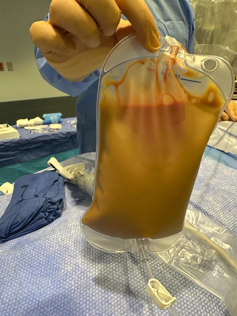
Case Presentation: A 37-year-old female with systemic lupus erythematosus (SLE), noncompliant with mycophenolate mofetil (MMF) and hydroxychloroquine, complicated by Class V membranous lupus nephritis and cirrhosis due to autoimmune hepatitis, presented with generalized weakness and altered mentation. This followed a recent admission for methicillin-sensitive Staphylococcus aureus (MSSA) bacteremia and acute kidney failure requiring hemodialysis (HD), during which she left against medical advice.On arrival, the patient was hypothermic, tachycardic, and hypotensive, necessitating vasopressors. Notable findings included lethargy, diffuse edema, and cool extremities. Labs revealed severe lactic acidosis (9.6 mmol/L), hemolytic anemia (total bilirubin 9.8 mg/dL), thrombocytopenia, hypoglycemia, elevated B-type natriuretic peptide, elevated ANA, and low complement levels. Imaging identified a large pericardial effusion with mass effect on the cardiac chambers, consistent with tamponade. She underwent emergent pericardiocentesis with 1.35 L of purulent fluid drained, which grew MSSA on culture, confirming purulent pericarditis. The patient’s HD catheter was removed, and she received a 6-week course of cefazolin (due to inability to rule out endocarditis).
Discussion: Purulent pericarditis, characterized by gross or microscopic purulence in the pericardial space, is rare in the post-antibiotic era. Staphylococcus species are the most common pathogens, accounting for 31% of cases. Mechanisms include hematogenous spread from bacteremia, direct intrathoracic extension, or myocardial involvement, such as infective endocarditis. A prior history of aseptic pericarditis may predispose to bacterial colonization, as seen in nearly half of cases in the post-antibiotic era. Cardiac tamponade is a common complication, with an incidence ranging from 42% to 77%. Diagnosis relies on pericardial fluid sampling, which also serves as source control. Antibiotic therapy typically spans 2–4 weeks.Arguably, patients with active lupus may be at high risk for purulent pericarditis. While the connection between SLE and purulent pericarditis is not well established, disease activity is a risk factor for bacterial infections in SLE, and sterile pericardial inflammation in active SLE may predispose patients to bacterial seeding. Serositis is a marker of active SLE, as 92% of patients with serositis have evidence of active disease in other systems, and 75% of patients with SLE have shown signs of active pericardial inflammation at time of autopsy. Although acute pericarditis is common in lupus, cardiac tamponade is rare (2% incidence). This case highlights the importance of considering purulent pericarditis in lupus patients presenting with tamponade, especially in the context of active disease and bacteremia.
Conclusions: This case underscores the need for heightened clinical suspicion of purulent pericarditis in patients with SLE who present with cardiac tamponade and bacterial infections. Further research is warranted to explore this association and improve management strategies.