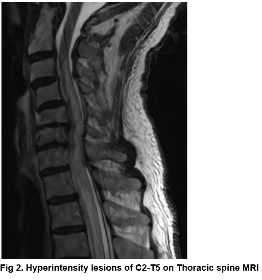
Case Presentation: An 88-year-old male with a past medical history of type 2 diabetes, hypertension, and hypothyroidism presented with acute onset right leg paralysis.The patient awoke with complete right leg paralysis, but otherwise asymptomatic and had normal mentation. Vital signs were unremarkable. Physical exam was notable for decreased sensation in the entire right lower extremity, decreased right patellar and Achilles tendon reflexes, and complete paralysis (0/5 strength). He had no rash. Initial labs revealed a BUN of 57 mg/dL (8-25 mg/dL), creatinine of 2.17 mg/dL (unknown baseline) and a Hg of 9.6 d/L (unknown baseline).A CT of the head without IV contrast showed age-related volume loss and microvascular ischemic changes, and was negative for hemorrhage or masses. An MRI of the brain, cervical and thoracic spine without IV contrast revealed severe T2/FLAIR hyperintensity throughout the cerebrospinal fluid (CSF) and extending from C2-T5 levels, with expansion of the cord parenchyma in the lower cervical and upper thoracic spine and mild inflammation of the optic nerve. Autoimmune processes such as neuromyelitis optica, were considered, however anti-aquaporin-4 antibodies were negative. Lumbar puncture revealed protein of 362 mg/dL (15-45 mg/dL), glucose of 97 mg/dL (40-75mg/dL), 10 red blood cells, and 42 nucleated cell (28% neutrophils, 51% typical lymphocytes), albumin of 269 mg/dL(8-42 mg/dL). Cytology was negative and no monoclonal bands were present. Varicella Zoster virus (VZV) PCR from the CSF was positive and the patient was diagnosed with VZV encephalomyelitis.Treatment was initiated with acyclovir 10mg/kg IV daily and methylprednisolone 1g IV daily. Repeat MRI showed resolution of the CSF enhancement, but the patient developed a progressive encephalopathy, autonomic dysfunction, and passed away.
Discussion: After primary infection, VZV remains latent in dorsal ganglia and cranial nerve until reactivation. Reactivation typically occurs in elderly or immunocompromised patients, presenting with a dermatomal vesicular rash that is painful and pruritic, although rash is not always present. Progression of VZV reactivation to encephalomyelitis is rare, and can present with focal neurologic deficits, headache, lethargy, visual deficit, hemiplegia, sensory deficits, urinary dysfunction, and coma. The incidence of VZV encephalitis is ~0.1-0.2%, with a mortality of 5-10%, and up to 80% for those who are immunocompromised. The finding of a rash can lead to a clinical diagnosis of VZV, or the diagnosis can be made with positive VZV in PCR testing of a skin lesion, saliva, or from the CSF.For patients with VZV encephalitis, MRI brain is typically normal or shows focal hyperintensity in the basal ganglia, thalami or cerebellum. This patient’s MRI showed diffuse CSF enhancement on T2 FLAIR, suggesting widespread disruption of the blood-brain barrier (BBB), a rare finding. Symptoms and disease progression may lag behind imaging, and as this case demonstrates, normalization of imaging does not equate with prognosis.
Conclusions: Encephalomyelitis is a rare complication of VZV reactivation. VZV can disrupt the blood-brain-barrier, demonstrated on MRI by T2/FLAIR hyperintensity, and clinical presentation may lag behind imaging findings. Early recognition of such imaging abnormalities is important for prognosis and can guide goals of care discussions.
.png)