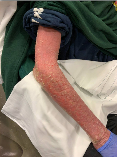
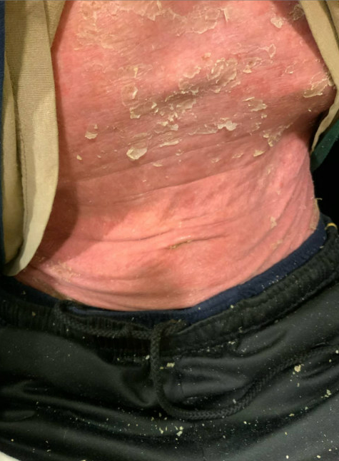
Case Presentation: A 62 year-old patient with stage III chronic kidney disease (baseline serum creatinine ~2.0 mg/dL), chronic obstructive pulmonary disease, and heart failure with preserved ejection fraction (HFpEF) was admitted from dermatology clinic with a painful body-wide rash. Two years prior to admission, he had a rash on the legs that improved with topical clobetasol and tacrolimus but was lost to follow-up. On the day of admission, he presented to dermatology clinic with erythroderma and unintentional weight loss of 40 lbs. and was directly admitted. He was afebrile with stable vital signs. On exam, he had confluent erythematous plaques with thick scale on the head, face, torso, hands, and upper & lower extremities comprising ~95% of total body surface area (Figure 1 and 2). Initial labs included hemoglobin 9.8 g/dL, platelets 683,000/mcl, no increased peripheral eosinophilia, serum creatinine 2.02 mg/dL, hepatic panel within reference ranges and lactate dehydrogenase 263 units/L. Two sets of peripheral blood cultures were positive for methicillin-resistant Staphylococcus aureus. Two punch biopsies were obtained. Vancomycin was started for MRSA bacteremia (presumed secondary to skin breakdown). TTE and repeat blood cultures were negative. He received intravenous (IV) fluids due to dehydration from erythroderma. He received 1 gm IV methylprednisolone later tapered to oral prednisone and topical triamcinolone and hydrocortisone with wet wraps. After one week, he developed dyspnea, hypoxemia (requiring 2-3 L supplemental oxygen), and peripheral/scrotal edema due to HFpEF exacerbation. Intravenous fluids were stopped and he received diuresis. Skin biopsies showed subacute to chronic spongiotic/eczematous dermatitis. Peripheral blood flow cytometry showed CD4:CD8 ratio of ~10 with no specific T cell marker abnormalities or monotypic B cell populations. Immunohistochemistry and T cell rearrangement clonality was negative for cutaneous T cell lymphoma (CTCL).
Discussion: Erythroderma is an exfoliative dermatitis involving greater than 90% body surface area, that can be life-threatening. Erythroderma occurs due to many causes, including psoriasis, atopic dermatitis, drug reactions, malignancy (solid tumors and hematologic malignancy such as CTCL), infections (scabies, dermatophytosis, Staphylococcal and Streptococcal scalded skin syndrome). Malignancy must be ruled out. In this patient elevation in LDH and the CD4:CD8 ratio on flow cytometry was concerning for CTCL or paraneoplastic erythroderma. Early dermatology consultation is crucial, as skin biopsy should be done before starting steroids, which can affect diagnostic testing. Initial aggressive fluid resuscitation is key given extensive insensible fluid loss due to skin barrier compromise.1 Patients with cardiac or renal disease may not tolerate fluid resuscitation,2 as was seen with this patient. In this patient skin biopsy, flow cytometry and immunohistochemistry ruled out CTCL and malignancy associated erythroderma. The final diagnosis was severe atopic dermatitis leading to erythroderma. Erythroderma is rare, with an estimated incidence of 1 in 100,000 and of these cases 10-18% are due to severe atopic dermatitis.3,4
Conclusions: Erythroderma can be severe and life-threatening. Early dermatology involvement is necessary to determine the cause and start prompt treatment. Aggressive fluid resuscitation is necessary but can lead to iatrogenic complications in patients with underlying cardiopulmonary conditions.