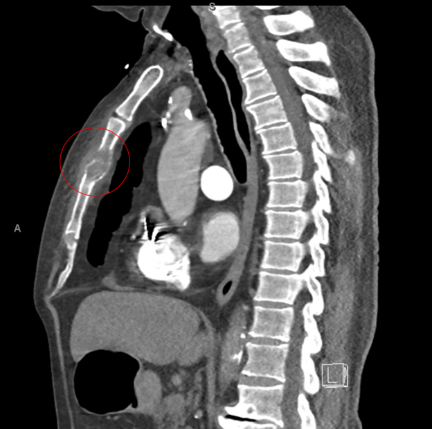
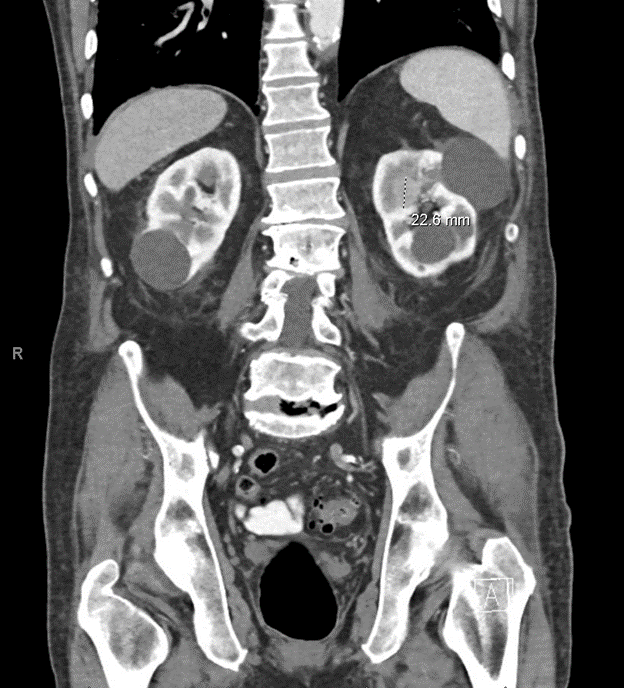
Case Presentation: An 83 year old male with past history significant for chronic obstructive pulmonary disease, active tobacco use disorder, essential hypertension, hyperlipidemia, complete heart block (status post permanent pacemaker), polycythemia, and thrombocytopenia (prior SPEP had shown two faint IgG lambda restrictions of unclear significance) presented to the emergency department with chest pain after a fall. Pain was localized to the sternum and exacerbated by any movement of the thorax and arms, deep breathing, and palpation. Vital signs were within expected normal ranges. Exam revealed exquisite tenderness to palpation of the sternum. Initial diagnostics included white blood cell count 11.72 x10^3/mcl, hemoglobin 16.3 g/dL, platelets 95 x10^3/mcl , sodium 135 mMol/L, blood urea nitrogen 54 mg/dL, serum creatinine 1.85 mg/dL, troponin-T 99 ng/L, creatine kinase 992 unit/L. A CT angiogram of the chest obtained to assess for pulmonary embolism showed an unexpected pathologic sternal fracture from presumed metastatic lesions as well as a presumed manubrial metastasis (Figure 1, red circle indicates sternal lesion). Additional diagnostics were obtained including serum protein electrophoresis (the two prior IgG lambda restrictions were unchanged), normal urine protein electrophoresis, normal serum free light chain ratio, and prostate specific antigen elevated at 7.05 ng/mL. A CT scan of the abdomen and pelvis showed a solid left renal mass in the parapelvic left upper pole of the kidney measuring 1.9 x 2.3 x 2.4 cm and several renal cysts (Figure 2); no lymphadenopathy was visualized. Biopsy of the sternal lesion showed squamoid/transitional appearance and immunohistochemistry positive for GATA3+, CK7+, CK20+, all supporting urothelial carcinoma (UCC) as the primary malignancy. Oncology was consulted and given poor prognosis, the patient received a single fraction of palliative radiation and discharged home on hospice.
Discussion: This case illustrates an uncommon presentation of metastatic UCC manifesting as pathologic sternal fracture. Isolated bone metastases from metastatic UCC are rare. In one study,1 only 6.7% of patients with metastatic disease have isolated bone lesions (as was the case in our patient). To our knowledge, there is only one other case in the literature2 of UCC metastatic to the sternum, which occurred with metastases to other sites. Our patient had metastases limited to the sternum. Initially, we considered multiple myeloma most likely, given pre-existing thrombocytopenia and acute kidney injury on presentation (with the osseous lesion this fulfilled two out of four “CRAB” criteria for multiple myeloma). However, on CT abdomen and pelvis, radiology felt the differential was most likely UCC due to the parapelvic location of the renal lesion, versus an atypical renal cell carcinoma (which are most often cortically located). As is typical, biopsy of a presumed metastatic site was crucial in diagnosing metastatic UCC. Given poor prognosis, this information facilitated rapid transition home on hospice.
Conclusions: When malignancy is on the differential, common sites of metastatic spread should always be prioritized but atypical presentations cannot be ruled out until definitive pathology is obtained. In this patient, the differential diagnosis evolved from multiple myeloma to renal cell carcinoma to urothelial cell carcinoma.