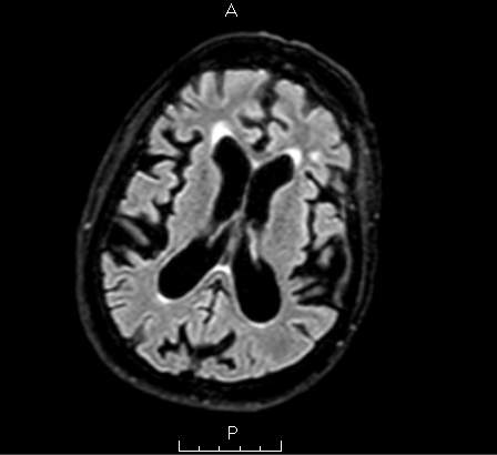
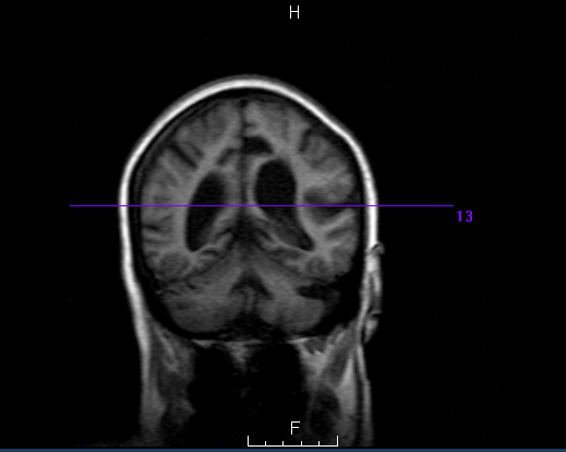
Case Presentation: A 50-year-old female with alcohol use disorder, asthma/chronic obstructive pulmonary disease (COPD), hypertension, and recurrent alcohol-related falls presented with generalized weakness, fatigue, and a recent fall striking her head. She denied neck pain but reported progressive imbalance and tiredness. The patient admitted to binge drinking for several days before presentation and stated that falls usually occur when intoxicated. Home medications included lisinopril 20 mg, loratadine, nicotine replacement, Symbicort, and Ventolin.On arrival, vitals were T 98.4 °F, HR 105 bpm, RR 18, BP 165/103 mmHg, and SpO₂ 98%. She appeared tired but alert, with mild expiratory wheezing, a right cheek abrasion, and mild abdominal distension without tenderness. Gait was ataxic without focal deficits.Laboratory results showed hyponatremia (Na 124 mEq/L), hypomagnesemia (1.6 mg/dL), and elevated ethanol (467 mg/dL). Other results were normal. She received 1 L IV normal saline, 2 g IV magnesium, thiamine, folate, ondansetron, and famotidine. Blood pressure was managed with lisinopril.CT head revealed mild-to-moderate ventriculomegaly out of proportion to sulcal widening, raising concern for normal pressure hydrocephalus (NPH). MRI brain showed moderate cortical atrophy, central volume loss, and periventricular small-vessel ischemic changes, suggesting possible alcohol-related atrophy or chronic microvascular disease. During evaluation, the patient left against medical advice (AMA) before further testing, including lumbar puncture and gait assessment. Follow-up attempts were unsuccessful, leaving the diagnosis undetermined.
Discussion: This case illustrates the diagnostic challenges encountered in evaluating gait instability, cognitive slowing, and recurrent falls in a patient with long-standing alcohol use disorder. The imaging findings raised concern for NPH, a potentially reversible cause of gait disturbance, yet were confounded by overlapping features of alcohol-related cortical atrophy and small-vessel ischemic disease. Metabolic abnormalities include hyponatremia, hypomagnesemia, and acute intoxication, which further complicated interpretation of the patient’s presentation, as each can contribute to ataxia and weakness.Distinguishing true NPH from hydrocephalus ex vacuo or alcohol-induced brain atrophy requires careful clinical correlation, including gait evaluation, cognitive assessment, and large-volume lumbar puncture; none of which could be completed due to the patient’s AMA departure. This underscores a critical hospital medicine issue: how social factors, substance use, and behavioral barriers can limit diagnostic completion, continuity of care, and patient safety.
Conclusions: This case emphasizes the importance of integrating imaging, metabolic findings, and psychosocial context in evaluating neurologic disorders in hospitalized patients. It highlights the complexity of diagnosing NPH amid confounding factors such as alcohol-related brain injury and small-vessel ischemia. The patient’s premature AMA departure prevented diagnostic confirmation, reinforcing the need for multidisciplinary strategies including early engagement, motivational interviewing, and social work involvement to mitigate the risks of diagnostic uncertainty and poor follow-up in patients with substance use disorders.