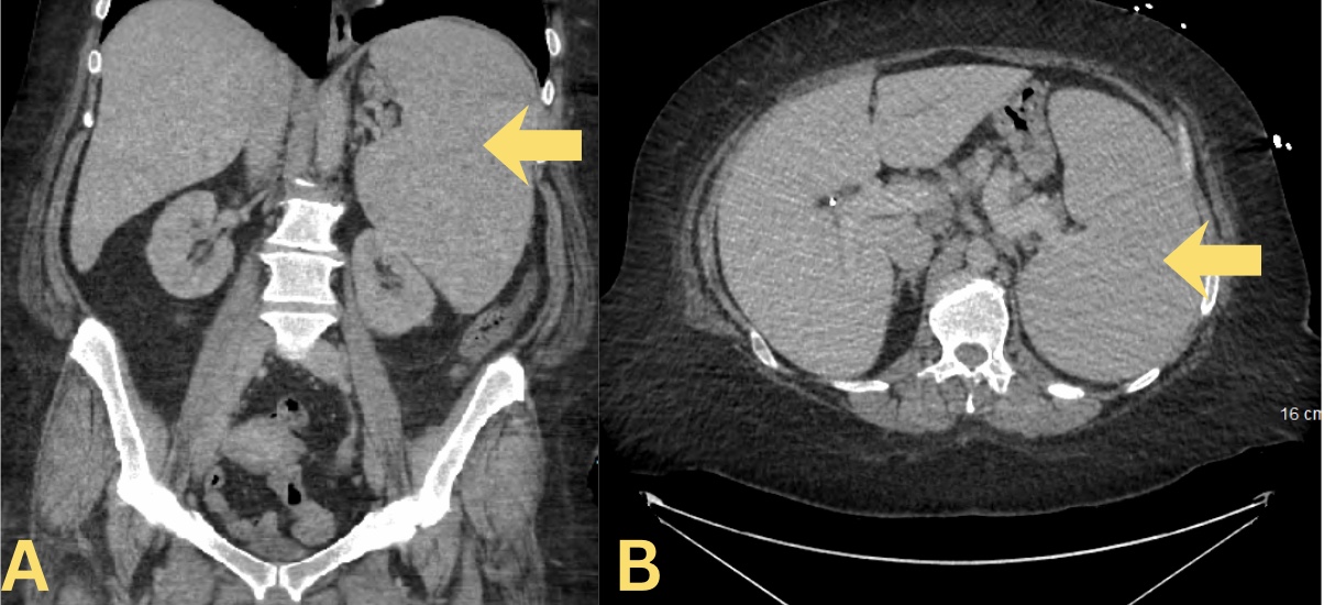
Case Presentation: A 45-year-old female presented with a complaint of painless jaundice worsening over 3-4 days, with chest heaviness, palpitations, and extreme fatigue. She also complained of intermittent numbness in all extremities over the past week. Her past medical history was significant for COPD, cigarette smoking, and chronic bilateral lower extremity swelling on diuretics. She was vitally stable on admission, and the physical exam showed scleral icterus and bilateral lower extremity edema. Blood work showed hemoglobin (Hb) of 5.2 gm/dL with macrocytosis (MCV 112.2 fL), reticulocytosis of 19.1%, haptoglobin < 10 mg/dL, and total bilirubin 3.83 mg/dL. Peripheral smear showed elliptocytes, teardrop cells, spherocytes, and large platelets. Serum immunofixation electrophoresis was significant for IgA monoclonal proteins with an elevated kappa light chain (22.4 mg/L) with no M spike. Serum protein electrophoresis and multiple myeloma panel came back negative. Direct antiglobulin test (DAT) was negative for IgG and positive for C3. CT abdomen and pelvis with contrast revealed massive splenomegaly of 21 cm with enlarged non-specific porta hepatic and retroperitoneal lymph nodes. Bone marrow biopsy showed no evidence of lymphoproliferative disorder. She got further short of breath in the middle of the night, and her Hb dropped to 4.3 gm/dL. She was administered 4 units of leukoreduced packed RBCs. 2 doses of 140 gm IVIg were given, in addition to IV methylprednisolone 40 mg twice daily during hospital stay. A diagnosis of cold agglutin disease (CAD) was made, and she was transitioned and discharged on oral prednisone 150 mg daily, in addition to trimethoprim/sulfamethoxazole and acyclovir prophylaxis. Eventually, the oral steroid dose was tapered off over the next 6 months to a 40 mg daily dose. She once presented with low back pain radiating down to her bilateral legs. Imaging revealed that she had developed bilateral femoral neck fractures without trauma, for which she had to undergo bilateral hip arthroplasty. Follow-up investigation revealed a high reticulocyte count of 10%, with DAT negative for IgG and positive for C3. She was started on combination chemo-immunotherapy with bendamustine 90 mg/m2 and rituximab 375 mg/m2 for a total of 4 cycles. The patient completed the regimen with minor side effects, which were resolved on completion of therapy.
Discussion: CAD is a complement-mediated hemolytic anemia most often caused by monoclonal IgM antibodies. DAT is positive for C3 complement & negative for IgG in CAD. Current evidence shows that monoclonal IgA-mediated CAD is exceedingly rare and follows a benign course, which is in contrast to our case and thus can be a diagnostic challenge when presented in an acute setting. CAD should not be treated with chronic corticosteroid therapy, as response rates are low and remission rates are high. Current evidence recommends the use of a short course of bendamustine-rituximab combination as an effective first-line option in patients requiring CAD therapy. Our patient’s clinical trajectory, the first known instance in which an IgA kappa paraprotein served as the pathogenic trigger for cold agglutinin disease in a middle-aged female, aligns with this contemporary therapeutic principle.
Conclusions: This case highlights the role of the hospitalist in managing acute, severe hemolysis due to a rare form of CAD, including atypical IgA-mediated CAD. Early recognition, transfusion support, and initiation of correct therapy are a critical part of preventing morbidity.