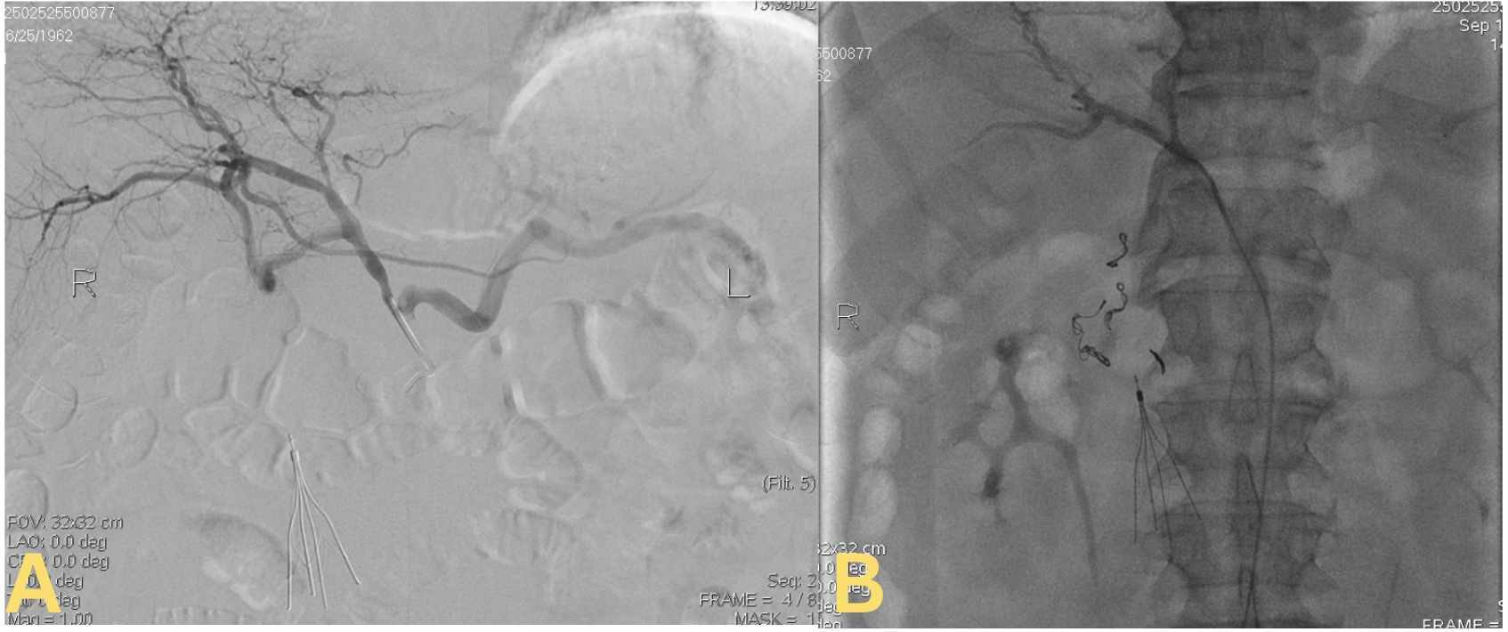
Case Presentation: A 63-year-old male presented with complaints of progressive shortness of breath and palpitations for three weeks, in addition to melena for the two days and an episode of coffee-ground emesis. He had been taking multiple ibuprofen tablets for the last 6 weeks for the lower extremity pains. He was tachycardic on arrival, but vitally stable. Labs showed hemoglobin 7.5 gm/dL, WBC count 37,000/μL, creatinine 1.32 mg/dL, blood urea nitrogen 26 mg/dL, and lactic acid 4.6 mmol/L. CT angiogram of chest revealed right middle and bilateral lower lobar pulmonary embolism (PE). Bilateral lower extremity Doppler was negative. He started on a heparin drip. In view of an ongoing active gastrointestinal bleed with hemoglobin dropping to 6.6 gm/dL, intravenous pantoprazole gtt was started, and heparin drip was held. He received 2 units of packed RBCs and underwent an IVC filter placement. The patient underwent esophagogastroduodenoscopy (EGD), which revealed a 22-mm non-bleeding cratered ulcer in duodenal bulb.Due to high risk of rebleeding, given the need for anticoagulation due to underlying PE, it was decided to perform prophylactic gastroduodenal artery (GDA) embolization. The celiac angiogram revealed a duplicate GDA, communicating with the superior mesenteric artery. Then, embolization of the medial branch was performed first, followed by the lateral one, with a total of five steel coils measuring 6.7 mm in size. Complete stasis of the GDA was confirmed angiographically. However, due to ongoing melena and worsening Hb, he was subjected to a repeat EGD, revealing an obstructing non-bleeding duodenal ulcer with no stigmata of recent hemorrhage, which was not biopsied in view of the patient’s anticoagulation status. A CT angiogram of abdomen demonstrated an ulcer in the first part of the duodenum with a segmental splenic infarct. It was decided to hold anticoagulation for 1 week for a possible biopsy to rule out the malignant nature of the ulcer, since the patient already had an IVC filter for interim coverage. The third EGD revealed a 15-mm wide-based, fibrotic, non-bleeding cratered ulcer in the duodenal bulb, with edge biopsies revealing benign pathology. The patient was ultimately discharged on warfarin and is currently doing well with regular INR monitoring.
Discussion: GDA, one of the two terminal branches of the common hepatic artery, is located directly behind the posterior duodenal wall. Due to its anatomical proximity, GDA is very commonly implicated in bleeding from duodenal ulcers, about 1.3-2.3 times more commonly compared to gastric ulcers. [1] To our best knowledge, it is the 3rd case of a duplicate GDA causing recurrent bleeding reported in the literature. In the previous two cases, Shah et al. recorded a case wherein duplicated GDA branches were embolized using 4 Tornado micro-coils, while Castater et al. chronicled a patient needing emergent duplicate GDA embolization with Gelfoam coil. [2-3]
Conclusions: Occurrence of duplicate GDA is increasingly rare but sometimes associated with increased risk of re-bleeding from duodenal ulcers. Life-threatening upper gastrointestinal bleeding from a posterior duodenal ulcer is managed with endoscopic and/or interventional treatment. Standard embolization with coiling might fail in rare variants of duplicate GDA, requiring thorough angiographic mapping to identify and embolize all feeding branches to achieve hemostasis. Duplicate GDA must be considered in recurrent duodenal ulcer bleed, with early interventional radiology involvement being the key.