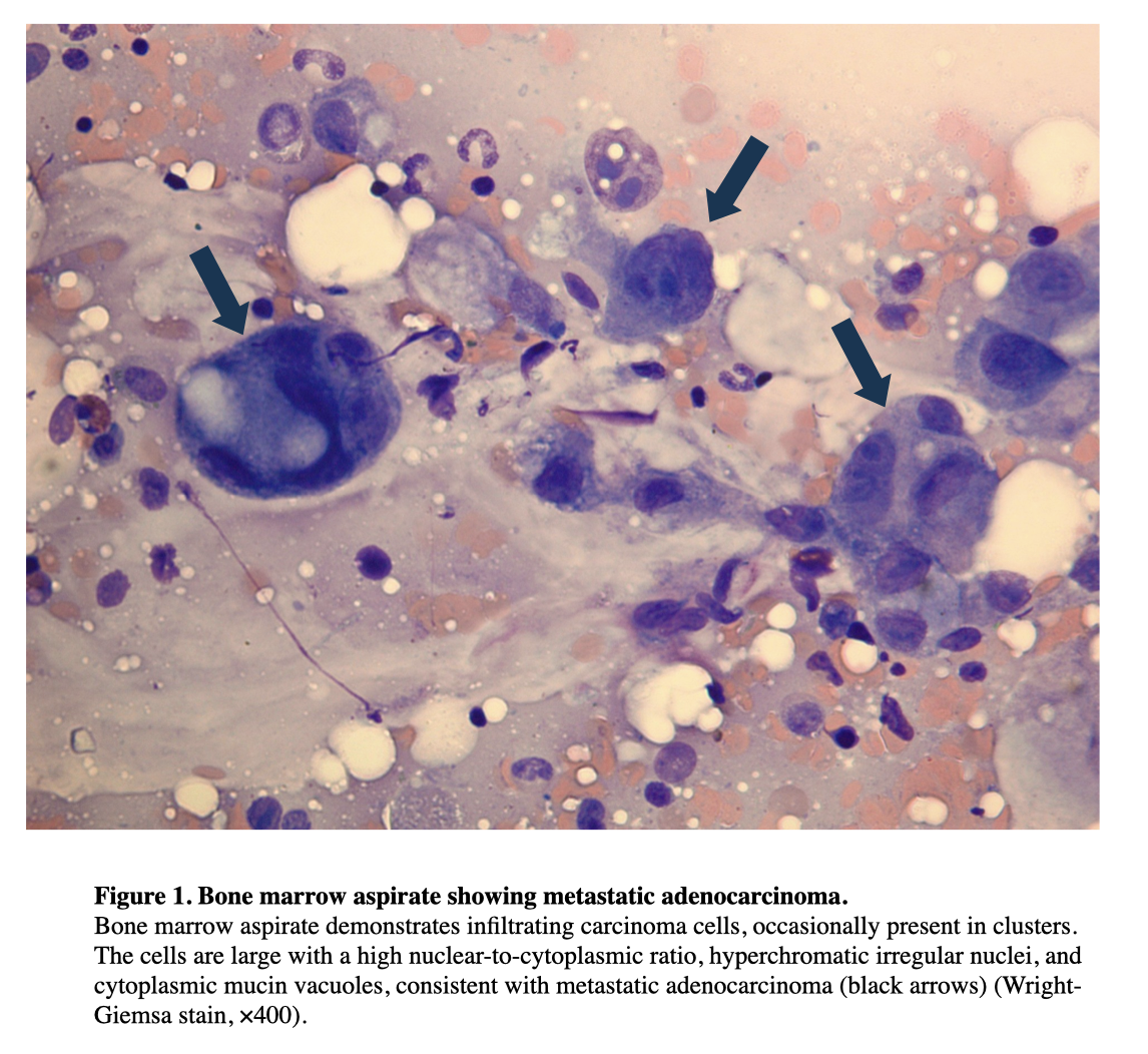
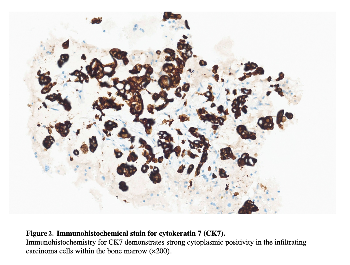
Case Presentation: A 50-year-old female with a history of stage IV metastatic lung adenocarcinoma who was initially treated with pembrolizumab, pemetrexed, carboplatin, and radiation for bony pain presented with weakness and shortness of breath and was found to have hemorrhagic shock requiring multiple blood and platelet transfusions. During hospitalization thrombocytopenia was refractory to high dose steroids, IVIG, and Nplate (Romiplostim). Despite receiving transfusions almost daily, she continued to have persistent anemia with no identifiable source of bleeding. Workup showed severe anemia to 4.0, thrombocytopenia to 13, elevated reticulocyte count, elevated LDH, very low haptoglobin, elevated unconjugated bilirubin and schistocytes on peripheral smear. Due to her refractory anemia and thrombocytopenia, TMA was suspected1. A bone marrow biopsy was then performed which demonstrated metastatic adenocarcinoma of mucinous type, confirming cancer-associated TMA. The patient also had melena which was thought to be indicative of GI end-organ damage, a known GI manifestation of TMA2. Despite restarting chemotherapy with carboplatin/paclitaxel, the patient continued to deteriorate with bacteremia and severe cytopenia and ultimately transitioned to comfort measures only.
Discussion: Thrombotic microangiopathy (TMA) is a rare cancer-associated coagulopathy that can either be caused by the malignancy itself or from treatment. Microangiopathic hemolytic anemia (MAHA) with thrombocytopenia due to thrombus formation suggests TMA. In cancer-associated TMA, it is thought that metastatic tumor cells induce a prothrombotic state1. In contrast, therapy-associated TMA may drive endothelial damage that leads to MAHA and thrombocytopenia, ultimately causing end organ damage1. TMA is increasingly associated with tumors that are mucin-secreting or cause paraneoplastic syndromes. This may be the first presentation of malignancy or can be seen in more advanced disease. Despite its rarity, TMA in oncology patients is a potentially fatal condition with high morbidity, mortality, and poor prognosis1. A high index of suspicion is therefore crucial for prompt recognition and treatment. TMA occurs in patients with various malignancies, the most common cases being gastric carcinoma, followed by breast cancer, prostate cancer, and lung cancer3. There are currently no guidelines to determine the diagnosis of TMA, however TTP, DIC, and atypical HUS must be excluded to avoid potential complications of therapeutic plasma exchange (PLEX). In patients with MAHA of unclear etiology, it is necessary to include a diagnostic bone marrow workup4. Management is usually supportive and focuses on treating the underlying malignancy or stopping the offending drug. Chemotherapy has been associated with improved survival in cancer-associated TMA5, but many patients have an extremely limited prognosis, with nearly a 50% mortality rate within one month of diagnosis6. Although this patient’s adenocarcinoma was no longer responding to her initial chemotherapy treatment at that time, the patient’s rapid decline was not only due to progression of her cancer, but also due to refractory TMA.
Conclusions: As evidenced by this case, it is important to recognize the symptoms of cancer-associated TMA in patients, which can be masked by cytopenia from cancer therapy or bone marrow infiltration. Early detection is imperative to guide appropriate management and potentially prevent rapid clinical deterioration.