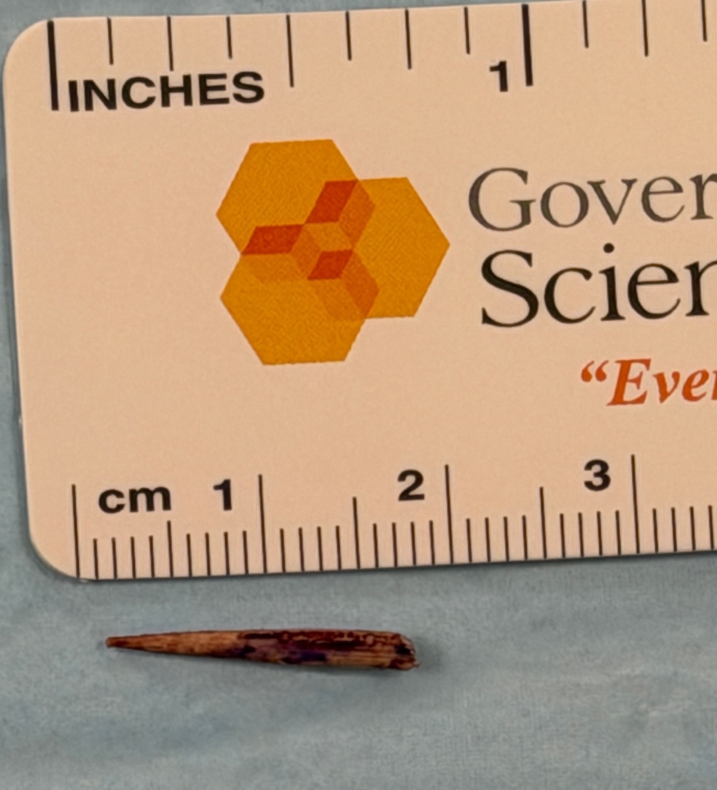
Case Presentation: An obese 69-year-old man with diabetes mellitus complicated by peripheral neuropathy presented with left foot and leg cellulitis. He recalled stepping on a sharp object while walking barefoot in his garage. He attempted dislodging the object with a flathead screwdriver. Three days later, he developed unbearable pain, swelling, and erythema which brought him to the emergency department.On physical examination, the left foot had striking erythema and 2+ pitting edema extending proximally to the tibial tuberosity. The left foot was tender to palpation, and focally intense erythema and swelling was present around the second toe. On the plantar surface, a small 1.5 mm puncture wound without purulence or drainage was seen over the second metatarsophalangeal joint. Podiatry evaluated the wound and recommended no surgical intervention. A CT scan showed soft tissue swelling without evidence of osteomyelitis or retained foreign body. Hospital medicine was consulted to admit the patient for intravenous antibiotics. Upon admission, a point-of-care ultrasound exam of the left foot revealed significant soft tissue edema (“cobblestoning”). Placing the transducer over the puncture wound revealed a prominent vertical acoustic shadow suggestive of a retained foreign body (Figure 1). Since the CT scan was unrevealing, an MRI scan with specific instructions to evaluate the base of the second toe on the plantar surface for a retained foreign body was obtained; however, the MRI report stated, “No foreign body identified.”The patient was admitted to the hospital and given intravenous ampicillin/sulbactam. Given the concern for retained foreign body and limited improvement after 36 hours of antibiotics, podiatry performed a surgical exploration in the operating room. A 1.8 cm wooden toothpick was expressed and removed from the wound (Figure 2). The patient’s cellulitis subsequently improved, and he completed a 10-day course of oral antibiotics.
Discussion: Point-of-care ultrasound has unique utility in identifying small radiolucent foreign bodies, most often wood, thorns, plastic, and glass. Approximately 80% of foreign bodies can be detected by plain radiographs, but wooden foreign bodies are not seen on plain radiographs in 86% of patients (1). When clinical suspicion exists, other imaging modalities, usually CT and MRI scans, are ordered, but clinicians should consider using ultrasound for further evaluation. Ultrasound can reliably detect wooden foreign bodies, even as small as 2-2.5 mm, most notably when retained in superficial soft tissues (2,3,4,5). Due to wood’s echogenicity and distinctive shadowing, ultrasound is the preferred imaging modality for suspected wooden soft-tissue foreign bodies (6,7,8). In contrast, CT is not ideal for detecting wood, as it often mimics the density of surrounding soft tissues (6,7,8). MRI has low sensitivity for detecting retained wood foreign bodies, particularly small fragments with no associated inflammatory areas; however, it may be useful in certain cases, including chronic or deep-seated wood fragments (6,7,8,9).
Conclusions: When suspecting a non-metallic retained foreign body composed of wood, glass, or plastic, clinicians should consider performing a point-of-care ultrasound exam which has demonstrated high sensitivity compared to CT and MRI as an imaging modality. Ultimately, as seen in this case, ultrasound is an efficient and effective method for conducting a dynamic and targeted evaluation for a retained foreign body.
.png)