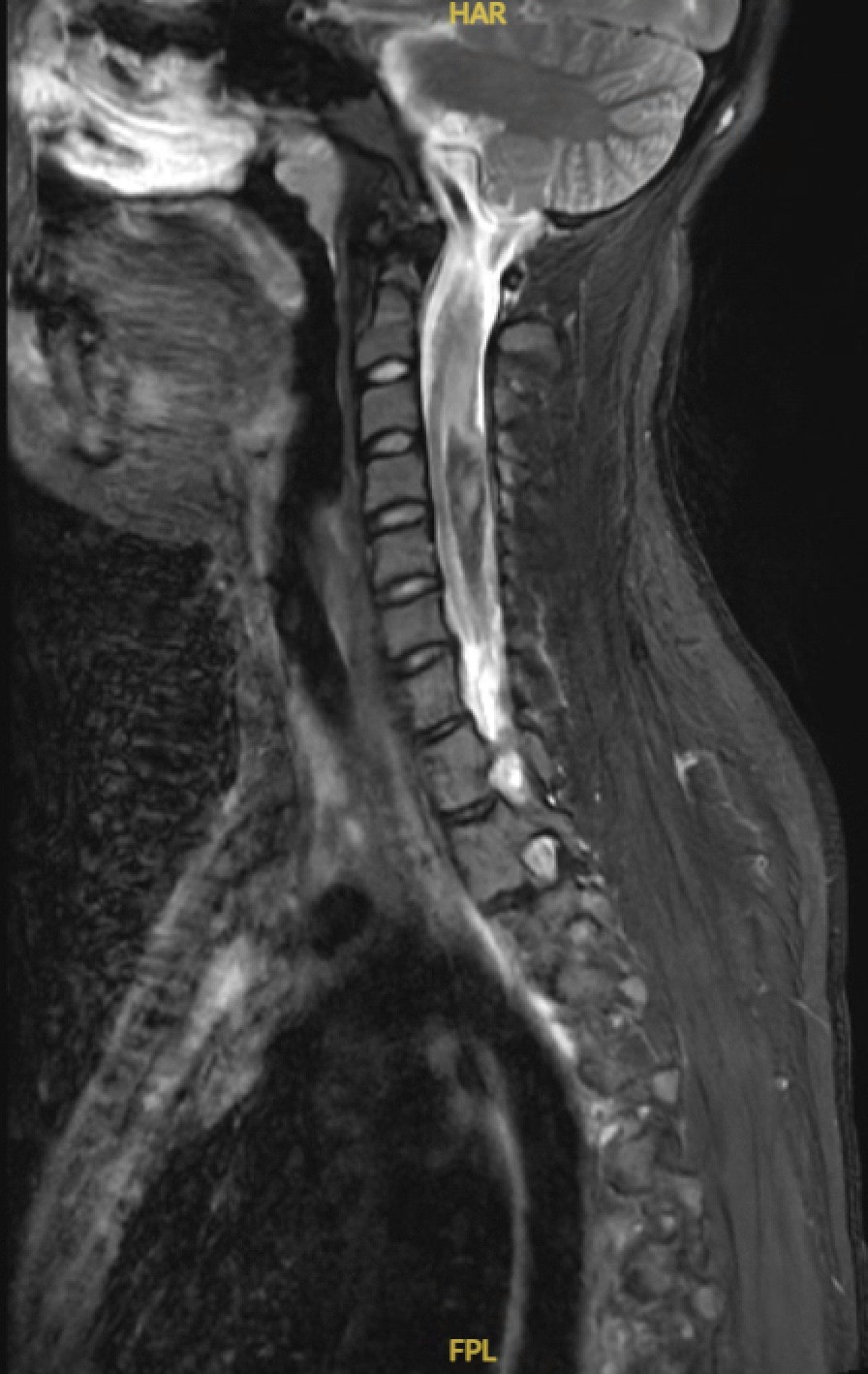
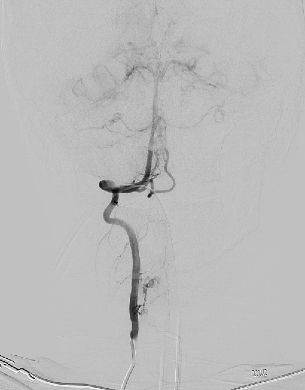
Case Presentation: 19 year old woman with a history of depression, anxiety, ADHD and pre-diabetes was admitted for evaluation of hypoxia and pneumonia. Two days prior she presented to an urgent care with sudden onset mid-back and right shoulder pain. She was diagnosed with muscle spasm and discharged with NSAIDs. The pain reoccurred the next day, prompting evaluation where she was found to be hypoxic. CT chest imaging demonstrated ground glass opacities. She was started on empiric antibiotics and transferred to our facility for treatment.Her exam was notable for inability to lift upper arms with grip intact. Examination suggested left foot drop with intact toe movement. Sensory testing showed decrease light touch sensation in bilateral lower extremities and absent patellar and ankle reflexes. She denied fall, trauma, bowel or bladder incontinence. Bladder scan showed significant urinary retention and digital rectal exam revealed no rectal tone. Emergent MRI spine and brain imaging ordered. MRI of the spine showed hemorrhagic lesions stretching roughly from C2-T6 with severe spinal cord compression. Emergent spinal angiography identified a ruptured cervical spinal cord AVM and associated cervicothoracic intramedullary hematoma. She underwent emergent C3-6 laminectomy with AVM resection, hematoma evacuation, and expansile duraplasty. Postoperatively, she developed tetraparesis and is currently undergoing intensive inpatient rehabilitation
Discussion: Spinal arterial vascular malformations (AVMs) are rare important causes of acute spinal cord dysfunction. Spinal cord infarctions account for approximately 1% of strokes, and hemorrhagic presentations represent only 3-4% of all AVMs [2]. These lesions can lead to neurological compromise through mechanisms such as mass effect, venous congestion, vascular steal, thrombosis, and hemorrhage. Early diagnosis is critical, as deterioration often occurs rapidly after onset.Hemorrhagic presentation in young patients without trauma are uncommon and frequently lead to diagnostic delays. Symptoms may be nonspecific—ranging from back pain to subtle motor or sensory changes—resulting in misdiagnosis [1]. Studies highlight that atypical or evolving symptoms in young patients should prompt neuroimaging to evaluate for spinal vascular pathology [3,5]. In this case, pain and hypoxia contributed to an anchoring bias toward pulmonary cause, highlighting the need for a broad differential and neurological assessment.Prompt recognition and intervention are essential. Neurological prognosis is closely tied to the time from symptom onset to diagnosis and surgical treatment. Management often includes microsurgical resection and/or endovascular embolization, with the goal to prevent rebleeding. Cure rates vary by AVM subtype and complexity, with the goal of complete obliteration to prevent rebleeding or further neurological decline. In a multicenter cohort of 463 patients, overall cure rate was 40.6%, with 58.5% after microsurgery and 26.4% after microembolization [4]
Conclusions: This case underscores the importance of maintaining a broad differential diagnosis when a seemingly unrelated admitting diagnosis is being pursued. It highlights the need for heightened clinical suspicion for spinal vascular malformations in young adults presenting with acute myelopathy. Early recognition with multidisciplinary individualized treatment strategies are critical to optimizing neurological recovery and minimizing morbidity.