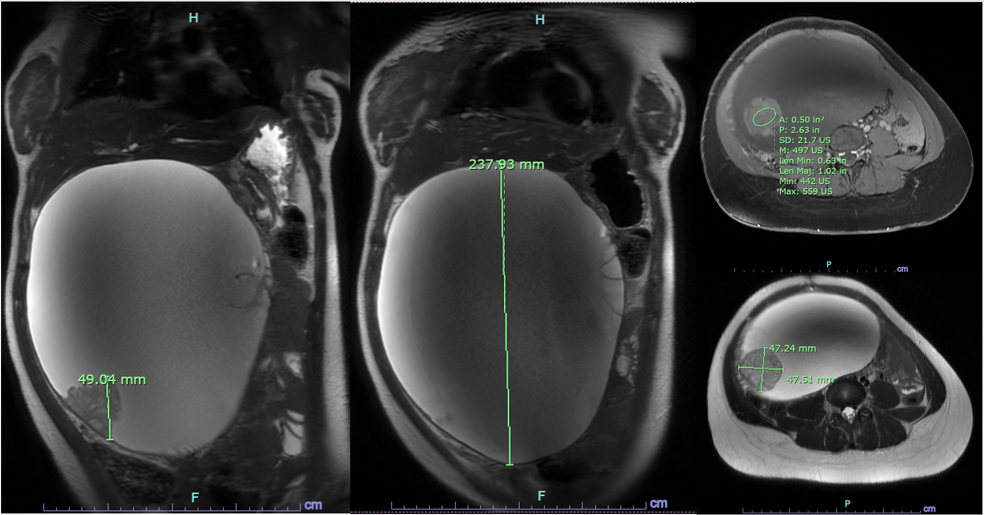
Case Presentation: A 14-year-old female with history of trisomy 21, hypothyroidism, chronic constipation, and recurrent ear infections presented for a right tympanostomy, during which she was noted to have abdominal distention. Her family reported progressive distension over the prior 2–3 months, which was attributed to constipation. Pediatric gastroenterology was consulted for suspected fecal impaction after an abdominal X-ray showed a paucity of bowel gas and moderate stool. She underwent a flexible sigmoidoscopy, with no fecal impaction noted. A nasogastric tube was placed and she was admitted for a bowel cleanout. Chest X-ray after nasogastric tube placement raised concern for a left upper quadrant mass, with recommendation for CT. A CT then revealed a 23.3 cm cystic mass with a 4.2 cm solid component displacing the right kidney, suggesting a retroperitoneal origin. MRI was limited due to intolerance of sedation but confirmed a complex cystic lesion. Tumor markers showed elevated CA-125 (46 U/mL) with normal CEA, AFP, estradiol, beta-hCG, and inhibin A/B. CBC and comprehensive metabolic panel were unremarkable. Repeat MRI under sedation suggested a right ovarian origin with mural nodules and mass effect, raising concern for malignancy. Pediatric surgery was consulted, and she underwent an exploratory laparotomy, which revealed a large right ovarian mass adherent to the fallopian tube. A right salpingo-oophorectomy was subsequently performed. Pathology confirmed a diagnosis of stage IA mucinous adenocarcinoma. Gynecologic oncology recommended genetic testing and ongoing surveillance.
Discussion: This case highlights a clinical scenario in which an unexpected, rare finding was obscured by features of a common pediatric complaint. In a patient with a history of trisomy 21 and chronic constipation, fecal impaction was the initial presumed diagnosis. However, persistent abdominal distention and careful re-examination of the imaging studies prompted further investigation, ultimately revealing an abdominal mass. When evaluating a pediatric patient with abdominal distention, common etiologies should be considered alongside less common, potentially life-threatening causes. Ovarian mucinous adenocarcinoma is rare, accounting for approximately 3% of epithelial ovarian cancers¹. However, around 65–80% of cases are diagnosed at an early stage, likely due to their typically large size¹. This patient’s history of developmental delay and constipation complicated her diagnosis. In cases of mucinous adenocarcinoma of the ovary, diagnosis at a later stage (III or IV) carries a median survival of 12–33 months¹. In contrast, stage I disease, as this patient had, has a 5-year survival rate exceeding 90%¹. Therefore, timely diagnosis and early detection can significantly influence outcomes.
Conclusions: Pediatric abdominal distention warrants a broad differential diagnosis, balancing common etiologies such as constipation with less typical but serious conditions, including malignancy. The physical examination can be especially informative in patients with developmental delay, where the history may be limited. Reassessment is essential to monitor the evolution of symptoms and avoid missing unexpected diagnoses.
.png)