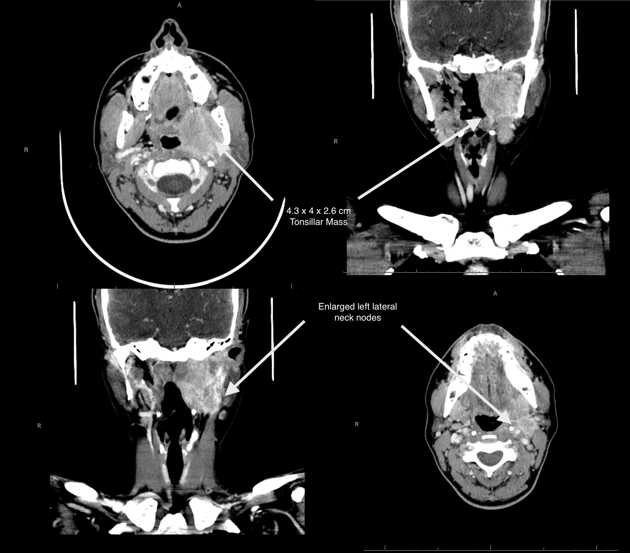
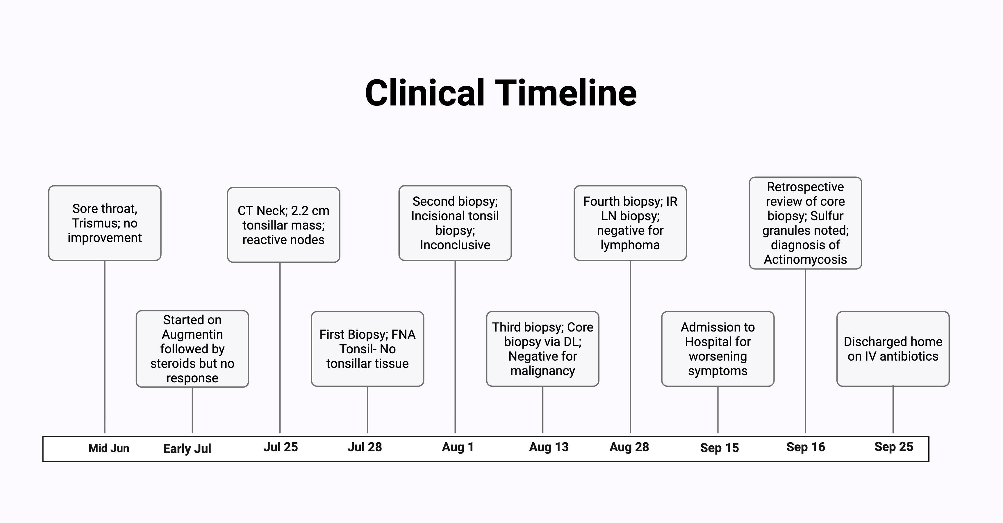
Case Presentation: Previously healthy woman in her early forties presented with persistent throat pain and a left tonsillar mass. Two months earlier, she had developed what seemed like a sore throat, but symptoms progressively worsened despite multiple antibiotic courses. The pain radiated to her left ear, was associated with jaw tightness, and trismus.She had been treated sequentially with Amoxicillin, Augmentin with steroids and Clindamycin without relief. Neck CT scan revealed a 2.2-cm left tonsillar phlegmon with reactive nodes highly suspicious for malignancy, prompting ENT evaluation. Due to her worsening odynophagia and weight loss, malignancy was strongly suspected, and she was referred to our cancer center.Her first and second tonsillar biopsies, performed five days apart, both showed benign squamous mucosa with nonspecific inflammation and bacterial overgrowth. A third core biopsy by laryngoscopy again showed reactive lymphoid hyperplasia and stromal fibrosis, but no malignancy. Ten days later, she presented to our center with persistent severe pain, trismus, dysphagia, and 12-lb weight loss.Labs showed leukocytosis (14.5 K/µL), anemia (Hgb 10.3 g/dL), and elevated inflammatory markers (CRP 82.6 mg/L, ESR 80 mm/hr). Repeat CT showed enlargement of the tonsillar mass to 4.3 × 4.0 × 2.6 cm with mild airway indentation. A fourth biopsy again negative for malignancy. Retrospective review of the prior third biopsy, sulfur granules were identified findings pathognomonic for actinomycosis.She was admitted for airway monitoring and started on broad-spectrum intravenous antibiotics, including piperacillin–tazobactam and daptomycin, with rapid clinical improvement.
Discussion: Tonsillar actinomycosis is rare and often misinterpreted as malignancy because of its firm, infiltrative appearance and poor response to standard antibiotics. Actinomyces are commensal, filamentous, gram-positive bacteria that become pathogenic when mucosal integrity is breached, typically after dental or tonsillar infection. The infection progresses slowly, forming fibrotic masses that mimic neoplastic lesions on imaging and examination.Similar presentations have been reported where repeated biopsies failed to show malignancy until sulfur granules were recognized on careful histopathologic review (1,2). In rare instances, the disease can be locally destructive and even fatal (3). Culture confirmation is difficult due to prior antibiotic exposure, making microscopic identification essential.Treatment involves prolonged high-dose penicillin or equivalent antibiotics for several weeks, occasionally requiring surgical drainage. Prompt recognition of actinomycosis can spare patients unnecessary surgery and distress when a unilateral tonsillar mass persists despite antibiotics and benign biopsies.
Conclusions: This case illustrates how tonsillar actinomycosis can closely mimic malignancy, leading to repeated invasive testing. Maintaining a high index of suspicion and carefully reviewing biopsy specimens for sulfur granules can prevent unnecessary procedures and anxiety. Early recognition and antibiotic therapy result in rapid recovery and excellent outcomes.