Case Presentation:
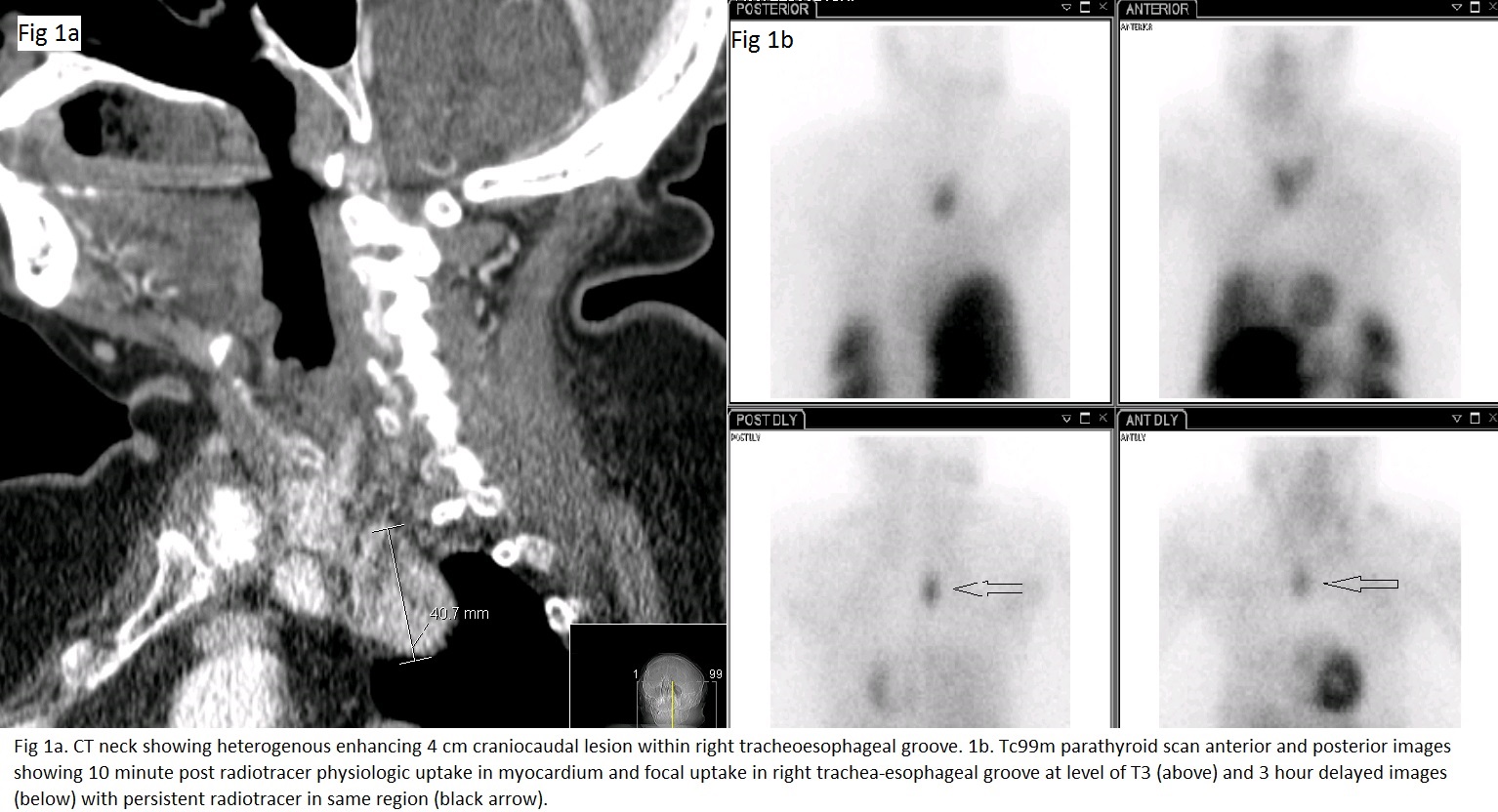
A 64 year old woman with a history of diabetes and hypertension presented to hospital with lethargy and disorientation to time, place and person. The vitals were: BP 232/127 mm Hg T 102.7°F PR 120 bpm RR 22. One week prior, she was diagnosed with a left otitis externa and received treatment with ciprodex ear drops. On this presentation, a CT head and temporal bone revealed opacification to the left mastoid and severe otomastoiditis with focal dehiscence. A left mastoidectomy with myringotomy was performed and the patient given broad spectrum antibiotics. Despite early management of her sepsis and surgical intervention, she was persistently somnolent requiring intubation for airway protection. Laboratory investigations revealed calcium of 14.6 mg/dl, Bun 38 mg/dl, creatinine 1.65 mg/dl, phosphorus 2.2 mg/dl. A PTH level was sent and found to be 720 pg/ml {range 10-55}. Following admission to the MICU ,treatment with saline diuresis, calcitonin and pamidronate, serum calcium level trended down. A concomitant improvement in mentation occurred, patient was stabilized and successfully extubated. CT neck revealed an enhancing lesion within the right tracheoesophageal groove, measuring 4 x 3.4 x 2.2 cm (fig 1a). A subsequent technetium 99m sestamibi scan showed an ectopic mediastinal parathyroid adenoma in the right trachea-esophageal groove at the level of T4 (fig 1b). Following improvement, patient was discharged with a plan for early outpatient surgery.
Discussion:
Parathyroid crisis is a serious and potentially fatal complication of primary hyperparathyroidism. Primary hyperaparathyroidism is due to a single adenoma in approximately 85% of cases and in 6-16% the adenoma is found in an ectopic site. Recent studies reported ectopic parathyroid glands were predominantly located in the thymus (38%), retro-esophageal area (31%), and within the thyroid (18%). Development of crisis is rare, occurring in approximately 1-2% of patients. Criteria postulated to define a crisis include: serum calcium level > 14 mg /dl, a markedly elevated PTH with signs and symptoms of profound hypercalcemia, including but not exclusive to altered mentation, renal dysfunction and gastrointestinal disturbances. Our patient was severely hypercalcemic with markedly elevated PTH, somnolence and acute kidney injury meeting criteria. Parathyroid crises usually occurs in response to a precipitating event, an otomastoiditis in this case, on the background of previously existent hyperparathyroidism. Aggressive medical management is an important bridge to urgent surgical intervention which is the definitive treatment.
Conclusions:
This was a case of a patient with an ectopic parathyroid adenoma who developed a parathyroid crisis in the setting of septic otomastoiditis. It highlights the need for a high clinical suspicion for hypercalcemia in the altered patient and an awareness that the presence of an infection can precipitate a life threatening crisis.