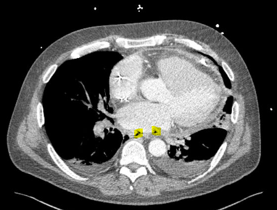
Case Presentation: A 62-year-old male with a history of paroxysmal atrial fibrillation presented with hypotension and 2 week history of chest pain after an ablation with pulmonary vein isolation. Transthoracic echocardiography demonstrated a pericardial effusion. Pericardial fluid sampling revealed an elevated leukocyte count of 130,000 cells per cubic millimeter and culture grew rare strep mitis/oralis and beta strep constellatus. While in the intensive care unit for vasopressor support, he developed left facial droop and hemiparesis, which spontaneously resolved within 1-2 hours. Initial non-contrast computed tomography (CT) of his head was unremarkable. He later became acutely altered with anisocoria and decerebrate posturing and repeat CT head was notable for pneumatosis cerebrii, consistent with air embolism. Computed tomography angiography (CTA) of the chest showed foci of gas within the posterior pericardial space adjacent to the left atrium (Figure 1A) concerning for atrio-esophageal fistula (AEF). No frank blood was found in his pericardial drain or oro-gastric tube and his hemoglobin remained stable. Cardiothoracic surgery performed an exploratory thoracotomy and repaired a left AEF.
Discussion: Atrio-esophageal fistula is an extremely rare complication of radiofrequency ablation with mortality rates approaching 100%, largely due to a delay in diagnosis1. The nonspecific presentation of AEFs is a contributor to this delay. Providers expect patients with AEFs to present with hematemesis or melena, given the communication between the atrium and esophagus. However, studies have found hematemesis in fewer than half of these patients2, and it was notably absent in our patient. This case highlights the need to maintain a high degree of suspicion for this complication in the appropriate clinical context.
Canadian centers provide patients who have undergone an ablation with a pocket card outlining the vague symptoms suggestive of an AEF and instructions for providers to have a low threshold to contact an electrophysiologist and avoid esophageal instrumentation post-ablation. From a prevention standpoint, small, single-center studies have shown reduced incidence of esophageal injury when luminal esophageal temperature (LET) monitoring is employed during RF ablation3.
Conclusions: Atrio-esophageal fistulas remain a rare, but potentially fatal, complication of atrial fibrillation ablations1. Raising awareness for this complication among both providers and patients as well as improved procedural monitoring for esophageal injury could hasten recognition and improve outcomes2.
1. Nair KKM, et al. “Atrioesophageal fistula: a review.” JAfib. 2015; 8(3): 1331.
2. Chavez P, et al. “Atrioesophageal fistula following ablation procedures for atrial fibrillation: systematic review of case reports.” Open Heart. 2015; 10;2(1):1-9.
3. Singh SM, et al. “Esophageal injury and temperature monitoring during atrial fibrillation ablation.” Circ. Arrhythm Electrophysiol. 2008; 1: 162-8.