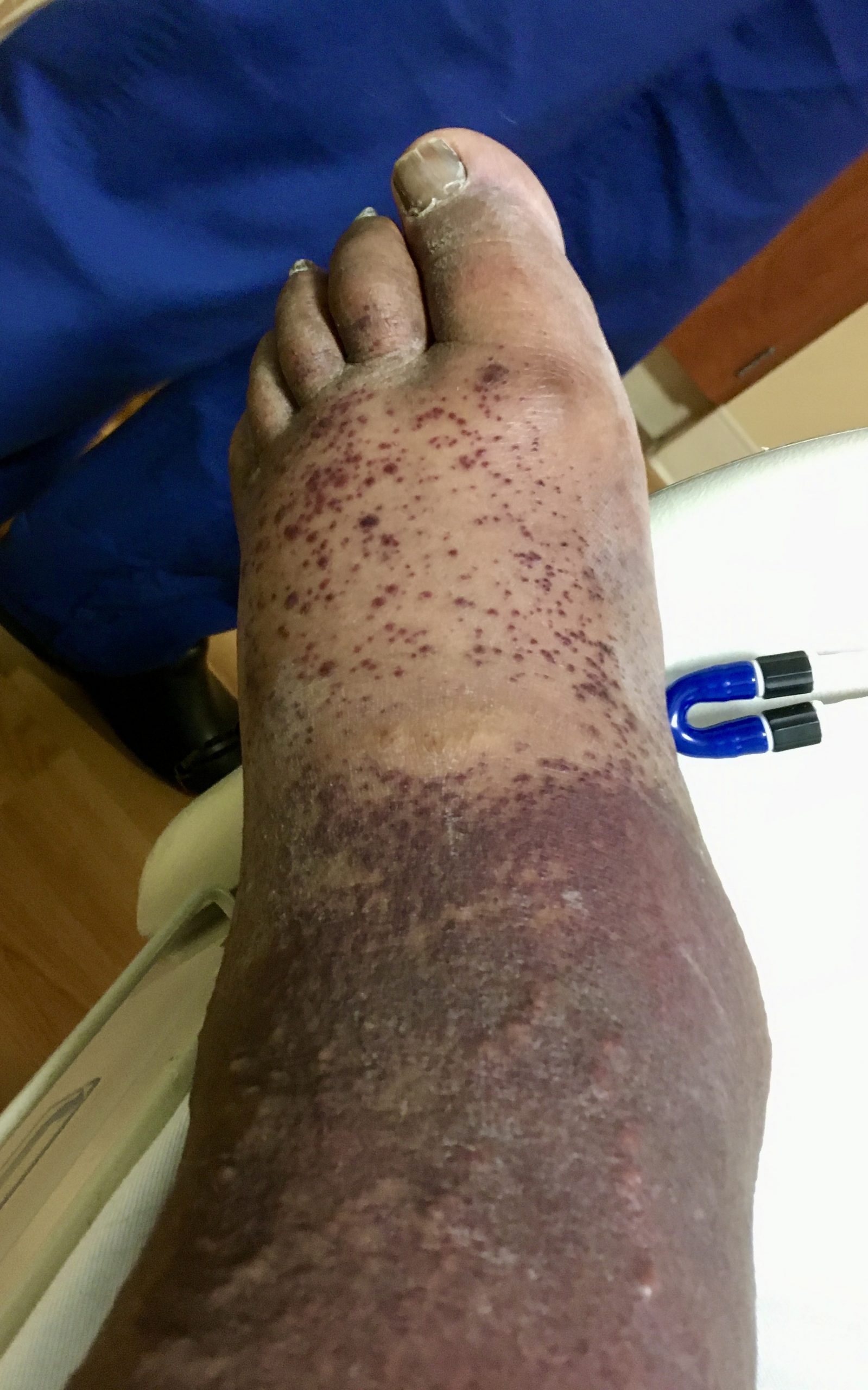
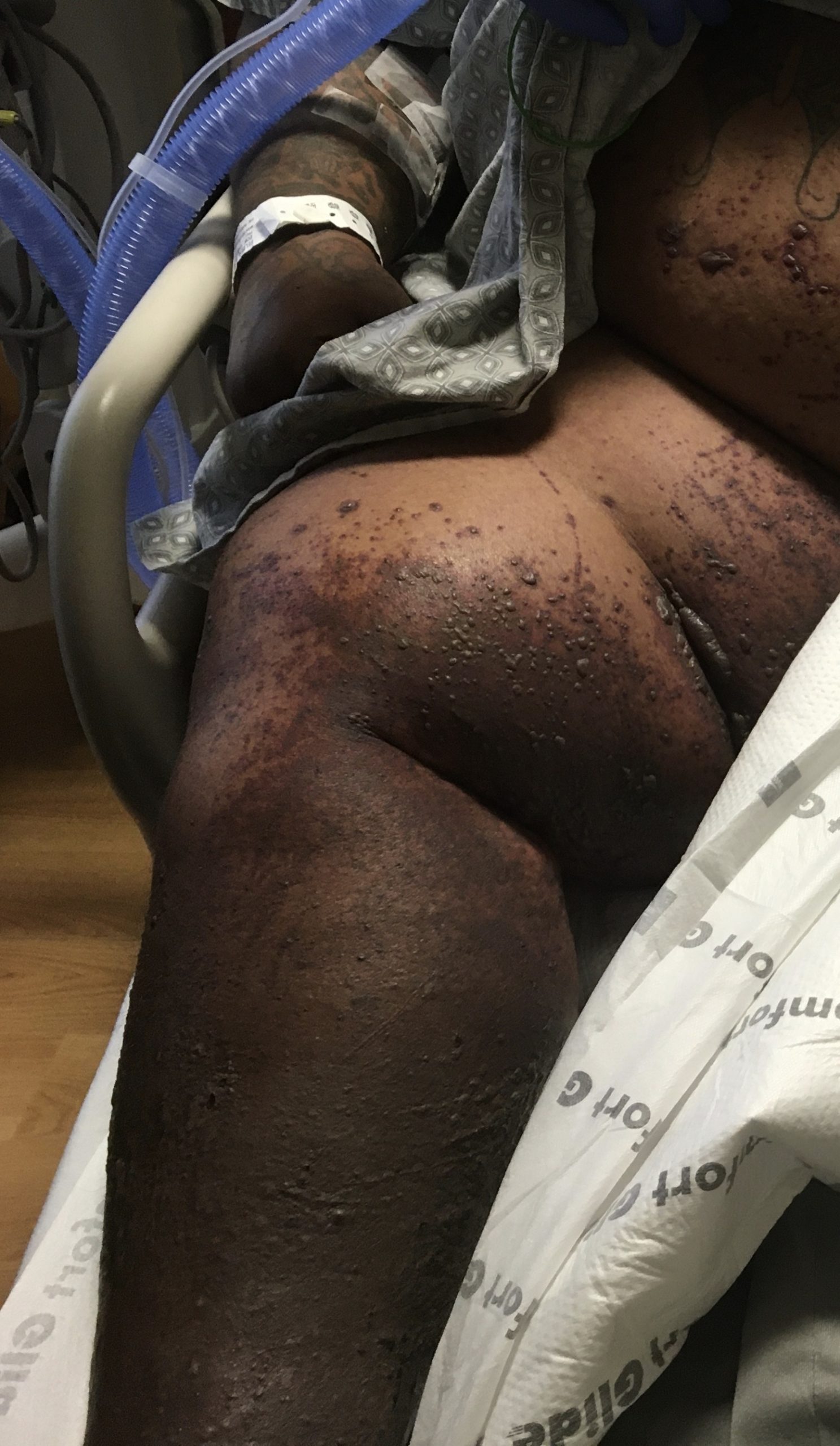
Case Presentation: A 48-year-old man with extreme obesity, heart failure, and a clinical diagnosis of gout presented with abdominal pain. Abdominal examination was limited by body habitus, as his body mass index was 68. Admission laboratory studies revealed acute kidney injury. He had been hospitalized before for similar presentations, but clear etiologies for the abdominal pain and acute kidney injury had not been found. On this occasion, acute tubular necrosis from either cardiorenal syndrome or prerenal azotemia topped the differential, but because the patient appeared euvolemic and there were no observed episodes of hypotension, the diagnosis remained in question.Shortly into the admission, the patient developed ankle pain and swelling, which was assumed to be a gout flare. The following day, an eruption of innumerable purpuric papules coalescing into plaques appeared over the dependent areas of his lower extremities and abdomen. The tetrad of abdominal pain, arthritis, renal dysfunction, and palpable purpura raised concern for IgA vasculitis. A skin biopsy confirmed the diagnosis when it revealed leukocytoclastic vasculitis with IgA-predominant immune complex deposition in the vessel walls. The patient’s renal dysfunction progressed rapidly, necessitating hemodialysis. Curiously, he also developed encephalopathy and hypoxia. Based on an arterial blood gas, hypoventilation from depressed alertness coupled with extreme obesity was suspected. However, although the hypercapnia corrected with noninvasive ventilation, the hypoxia did not. Chest radiograph revealed the development of bilateral hazy airspace opacities. Because volume status was optimal through the use of hemodialysis, pulmonary edema was unlikely. Infectious workup including sputum culture was negative. Only after receiving pulse-dose methylprednisolone, plasmapheresis, and mycophenolate mofetil did the patient’s respiratory status improve – along with his renal function, abdominal pain, and arthritis in concert. The existing purpura persisted but stopped proliferating.
Discussion: Pulmonary involvement is a rare manifestation of IgA vasculitis, occurring in an estimated 2.4 percent of cases according one Mayo Clinic study. This entity is described more in adults and most often presents as diffuse alveolar hemorrhage. It is hypothesized that the risk of developing pulmonary pathology increases with disease severity. This patient had persistent hypoxia even after correcting the mostly likely potential causes. Obesity hypoventilation syndrome coupled with encephalopathy seemed an obvious explanation, but he remained hypoxic even after the hypercapnia resolved with noninvasive ventilation. Were the patient not euvolemic, the bilateral hazy pulmonary opacities could have been ascribed to edema. Diffuse alveolar hemorrhage, however, would have a similar radiographic appearance. Unfortunately, the patient’s clinical instability and body habitus made him a poor candidate for advanced imaging or procedures, thereby curtailing further workup. In this context, a reasonable explanation for his persistent hypoxia is pulmonary IgA vasculitis.
Conclusions: Given the severity of this patient’s presentation, we suspect that this patient’s hypoxia, which persisted despite addressing other potential etiologies, was IgA vasculitis manifesting as pulmonary disease. Because vasculitides in general are rare, their diagnosis can be challenging. This case highlights a rare presentation of a rare vasculitis in adults.