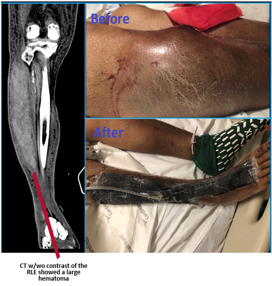
Case Presentation: A 57 year-old male with history of alcoholic cirrhosis, hypertension, and stage IV chronic kidney disease (CKD) presented to the hospital with bilateral lower extremity edema and shortness of breath. He was diagnosed with worsening CKD meeting criteria for hemodialysis, acute decompensated cirrhosis, severe sepsis due to spontaneous bacterial peritonitis complicated by pansensitive Klebsiella pneumoniae (KP) bacteremia. Despite appropriate treatment with piperacillin/tazobactam, leukocytosis persisted and ten days into hospitalization, he developed a lump on the right mid-shin, around a small ulcer. On physical exam, he was oriented to person only, tachycardic (120-130s) and tachypneic (mid 30s). Within an hour, he developed severe swelling on the right lower extremity (RLE) with acute bleeding from the ulcer not controlled with pressure. Emergency general surgery was consulted and placed 8 sutures around the ulcer and a pressure bandage was applied with minimal improvement. At the time of decompensation repeat labs showed an elevated PT/INR 22.4 and 1.87, platelets of 71,000 cells/cmm, hemoglobin of 4.4 g/dL (previously 7.8 g/dL), and WBCs of 17,000 cells/cmm. He received Desmopressin 0.3 mcg/kg to decrease the bleeding, one unit of plasma, and two of packed red blood cells. CT scan of the RLE showed a large hematoma extending from below the knee joint to the level of the distal tibial diaphysis measuring 26 cm x 5.8 cm x 6.5 cm, without evidence of infection or abscess in the underlying tissue. Due to persistent bleeding, the following day he underwent a surgical evacuation and was found to have an infected clot with purulent fluid positive for KP. He required a second excision and debridement and had a wound vacuum-assisted closure placed. On discharge to skilled nursing facility, his coagulation panel returned to baseline and creatinine improved, no longer meeting criteria for hemodialysis.
Discussion: Klebsiella pneumoniae is a gram-negative organism prevalent among immunocompromised middle to older age males including those with history of cirrhosis and alcoholism. It causes a wide range of infections but complicated skin and soft tissue infections of the LEs with community acquired KP are uncommon. Furthermore, monocrobial KP as the causative organism of a LE abscess is rare. Several cases of abscess formation due to KP have been reported in Taiwan, but few in the United States. The underlying abscess likely led to the hematoma formation as the patient suffered from an existing cirrhosis related coagulopathy and CKD related uremic platelet dysfunction. Differential diagnoses to consider for a hematoma should include intramuscular aneurysm or hemangioma, abscess, soft tissue malignancy or soft tissue carcinoma metastases.
Conclusions: Although CT scan is the best modality to diagnose an abscess, it was not diagnostic for this patient. Therefore it is very important to recognize that KP can cause a disseminated infection, particularly when treating a patient with persistent leukocytosis. KP can cause a seeded infection that if not treated in a timely manner can lead to increased mortality among the immunocompromised population.