Case Presentation: NA is a 24 yo African American female who presented with severe oral and genital ulcers with pain and inability to eat or speak. She started getting oral ulcers at age 4 with flares every 3 months, resolving quickly after prednisone. Her genital ulcers started at age 12, but were not as frequent as the oral ulcers. For 3 months prior to admission, she reported recurrent flares, being lesion free for only 1 week at a time. Her dermatologist switched her to cyclosporine 100 mg BID for 40 days believing this to be complex aphthosis with no biopsy. She reported fevers with flares but denied visual problems, joint pain, hair loss, dry eyes or mouth, abdominal or periorbital pain. She had no other past medical/surgical history and denied family history of lupus, RA, IBD, or autoimmune diseases.
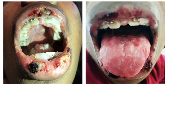
Physical exam revealed erosions with hemorrhagic crusting and fibrinous exudate on vermilion lips, dorsal, lateral, and ventral tongue, mucosal lips, hard and soft palate, and gingivae. Palms and soles were clear and she had normal genitalia with no ulcers or scarring. Serology was negative for ENA, anticardiolipin IgM/G, HSV-1/2 IgG, HIV, RPR, anti-centromere IgG, anti-Sm IgG, RNP IgG, Scl 70 IgG, SSA/RO and SSB/LA IgG, APS IgM/IgG, and no Herpes Simplex Virus (HSV) on direct fluorescent antibody test from the lip. Dermatology performed punch biopsies of mucosal lips for light microscopy and direct immunofluorescence showing erythema multiforme. She received 3 days of pulse IV methylprednisolone with marked improvement in her lesions and return to normal speech and oral intake by discharge.
Discussion: Erythema multiforme (EM) major is an immune-mediated condition characterized by cutaneous target lesions and mucosal membrane involvement that has a prevalence of less than 1%. Target lesions typically are seen with symmetric distribution on extensor surfaces of peripheral extremities. Mucosal lesions can involve oral, ocular, and/or genital mucosa and commonly present as vesiculo-bullous lesions rapidly developing into painful erosions. Histologic findings of EM are not specific, and mostly useful for excluding entities in the differential.
EM is generally considered to be an acute, self-limiting illness, but there is a subset of patients with EM who experience frequent episodes over many years who are diagnosed with recurrent EM. Recurrent EM has been linked to many different inciting factors, most commonly a preceding HSV infection. Other cases of recurrent EM have been linked to M. Pneumoniae, hepatitis C, menses, ingestion of benzoic acid, or have had no clear etiology.
Management for recurrent EM is treatment of the inciting factor, such as suppressive therapy for HSV. In severe forms of EM with functional impairment, early therapy with high dose steroids is considered, despite no controlled studies and controversy regarding risk of infections.
Conclusions: Our patient represents a unique case of EM due to the recurrent nature, no obvious inciting factors, no previous biopsies and no positive HSV. Ultimately, she had several recurrences with hospitalizations, received the final diagnosis after biopsy, and improved once treated. By identifying EM quickly, one can appropriately treat with steroids, both decreasing hospital workup and patient length of stay.