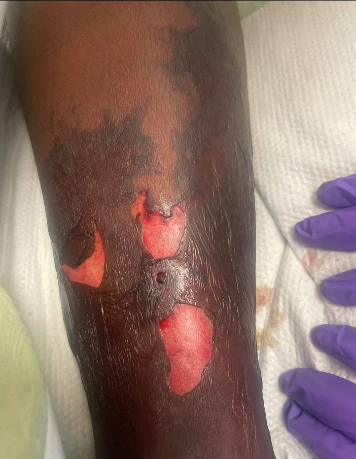
Case Presentation: A 43-year-old male with a past medical history of Atrial Fibrillation on Warfarin, Stage III Chronic Kidney Disease, and Chronic Systolic Heart failure (EF 15%) presented to the ED with diarrhea and dyspnea. He was found to be in Atrial fibrillation with Rapid Ventricular Response with CHF exacerbation and shock, and he was admitted to the ICU. The next day, the patient developed significantly altered mental status and continued to decompensate hemodynamically, eventually requiring cardioversion for RVR, urgent intubation, vasopressor support, and CRRT. Blood cultures showed Escherichia Coli (E. Coli) and enterobactereles, and the patient was empirically started on Cefepime and Vancomycin for sepsis. In the setting of diarrhea, a GI source was suspected, but workup was negative including stool C. diff, enteric panels, and abdominal imaging. On initial exam, the patient had left lower extremity erythema from above the knee to ankle, thought to be cellulitis. Through the next week, he developed left lower extremity retiform purpura with bullae that progressively worsened. Infectious Disease and Dermatology were consulted, and the patient had a skin biopsy, with cultures showing E. coli, and pathology showing “neutrophilic inflammatory infiltrate in the dermis and subcutis … and the degree of acute inflammation is suspicious for an infectious process”, thus confirming a diagnosis of Ecthyma gangrenosum (EG). Despite broad-spectrum antibiotic coverage and eventual resolution of bacteremia, the left lower extremity wounds continued to worsen. Workup for immunocompromising conditions including autoimmune diseases was normal. The patient ultimately needed surgical intervention with wound debridement twice and placement of a wound vacuum to promote healing. The patient was eventually discharged with plans to return for left lower extremity skin grafting at a future date.
Discussion: Ecthyma gangrenosum (EG) is a cutaneous manifestation often found on the gluteal or perineal regions or the extremities (1). Clinically, EG is described as erythematous nodules or vesicles that evolve into necrotic ulcers with eschar (2). P. aeruginosa accounts for 73% of infections associated with EG, but other bacteria have been seen, such as other Pseudomonas species, Staphylococcus, E. Coli, and Klebsiella pneumoniae (2). Although most often in the setting of bacteremia in immunocompromised patients, there has been more recent reporting of EG occurring in healthy individuals (2, 3). EG is treated with aggressive antibiotics and surgery if necessary, including skin grafting, although only 17.4% of EG cases require grafting (2).
Conclusions: As understanding of EG expands, cases of EG in immunocompetent patients have been recorded, but it remains a disease of primarily immunocompromised individuals. This patient had significant comorbidities, such as Chronic Kidney Disease and Chronic Systolic Heart Failure, that complicated his clinical course and contributed to his hemodynamic instability; however, he was immunocompetent on arrival. In addition, this case of EG resulted in severe extremity wounds that failed to improve with broad-spectrum antibiotics and required surgical treatment and skin grafting. This case of EG further emphasizes that a lack of immunocompromised state and P. aeruginosa infection are not grounds for eliminating EG in a differential diagnosis and demonstrates that multiple debridements and skin grafting are viable options to treat the disease.