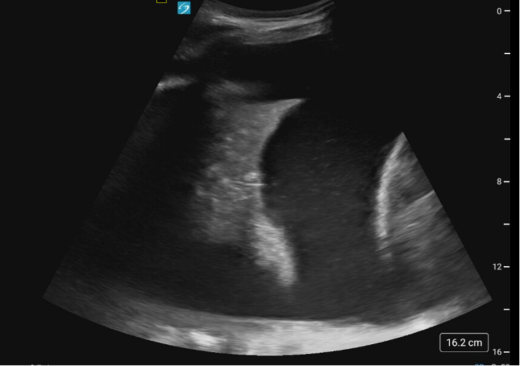
Case Presentation: A 41-year-old man with a history of cirrhosis secondary to primary sclerosing cholangitis and recurrent right hepatic hydrothorax (HH) presented to the emergency department (ED) with worsening dyspnea. The patient routinely underwent thoracenteses every 3-4 days as an outpatient and had missed his most recent thoracentesis. He had no prior history of spontaneous bacterial peritonitis (SBP). He was asymptomatic apart from his typical hydrothorax-related dyspnea.He was afebrile, pulse 101, blood pressure 163/92. He had diffuse jaundice and diminished breath sounds over right lower lung field. Bedside ultrasound showed no evidence of ascites but revealed a large simple right-sided pleural effusion (Figure 1). Laboratory workup was significant for a white blood cell count of 24.4 cells/μL, sodium 133 mmol/L, albumin 2.1 g/dL, and normal creatinine. Thoracentesis was performed with drainage of 2.5 L of serous fluid. Pleural fluid analysis showed 819 nucleated cells (87% neutrophils), LDH 56 U/L, glucose 151 mg/dL, and protein < 2.0 g/dL, consistent with a diagnosis of spontaneous bacterial empyema (SBE). Pleural fluid culture had no growth. The patient completed a total of a 7-day course of ceftriaxone, with repeat thoracentesis performed one day after completion of the antibiotic course showing improvement in pleural fluid cell count to < 250 nucleated cells.
Discussion: Hospitalists should be aware of SBE, which arises from bacterial seeding of a HH in a mechanism similar to that of SBP with ascites. A diagnosis of SBE is established by a pleural fluid absolute neutrophil count >250/mm3 or a positive pleural fluid culture.1 The term “empyema” is a misnomer, as SBE typically presents with a transudative rather than exudative effusion.2 SBE is a morbid condition, with treated mortality of 20-38%.3Our case is unusual in that the patient presented with SBE in the absence of ascites. SBE itself is relatively uncommon, occurring in only ~15% of cirrhotic patients with HH.3 Furthermore, of these patients with SBE, less than 10% of them will present with isolated HH without ascites.3 The mechanism by which HH develops in the absence of ascites is not fully understood. In addition to the positive pleural-peritoneal pressure gradient, it is postulated that patients with HH have peritoneal fluid reabsorption that outpaces pleural fluid reabsorption, possibly due to impaired lymphatic drainage at the diaphragm.4 As seen in our patient, up to a third of patients with SBE will present without infectious symptoms. Given the high mortality conferred by SBE, timely pleural fluid studies are imperative in patients with HH, even those who are asymptomatic or who present for regularly scheduled thoracenteses. A third-generation cephalosporin is generally sufficient empiric coverage for patients without risk factors for drug-resistant organisms, though appropriate response should be confirmed by repeat thoracentesis demonstrating reduction in pleural fluid cell count. Unlike the management of a parapneumonic effusion, chest tube placement is generally avoided in HH and SBE, as it can lead to volume depletion, severe electrolyte derangements, or fistula formation.2
Conclusions: SBE can uncommonly occur in cirrhotic patients in the absence of ascites. Given its high mortality, clinicians should maintain a high suspicion for SBE in patients with HH and promptly obtain pleural fluid studies, even in the absence of infectious symptoms.