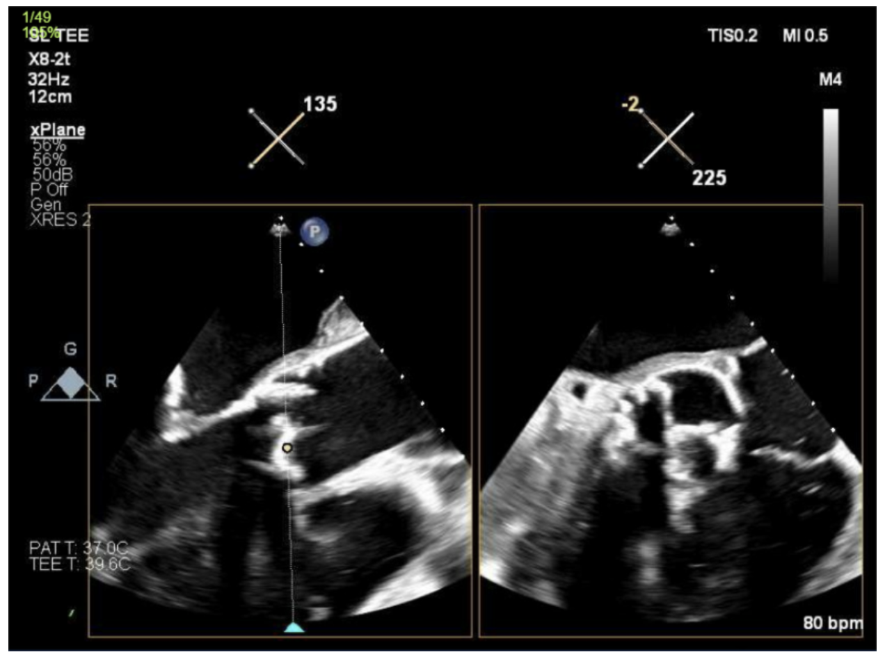
Case Presentation: A 35-year-old woman with end-stage renal disease (ESRD) on chronic hemodialysis, suspected drug-induced lupus with recurrent pericarditis, and severe bi-valvular disease requiring bioprosthetic aortic valve replacement seven months earlier, presented with progressive exertional dyspnea, early satiety, and worsening left upper quadrant abdominal pain initially thought to be a lupus flare. Laboratory evaluation was notable for elevated inflammatory markers. Initial CT abdomen was unrevealing however eventual repeat imaging of the abdomen demonstrated multiple wedge-shaped splenic infarcts concerning for an embolic process. Further workup revealed severe structural degeneration of the bioprosthetic aortic valve characterized by restricted leaflet mobility, heavy calcification, and an elevated transvalvular gradient, raising concern for catastrophic valve failure. Blood cultures were negative, and there was no clinical evidence of infective endocarditis. The patient was started on anticoagulation for presumed embolic events. Given the combination of progressive cardiac dysfunction, and the need for renal replacement therapy, she was deemed an appropriate candidate for heart-kidney transplantation.
Discussion: Thromboembolism is a recognized complication of bioprosthetic valve replacement, occurring at an estimated rate of 0.2–2.6% annually (1). Although the cerebral circulation accounts for most embolic events (75–80%), systemic embolization to visceral organs is far less common, with only 20–25% involving sites such as the spleen (2, 3). We describe a presentation of splenic infarction as the initial manifestation of bioprosthetic aortic valve degeneration in a young woman with multiple comorbidities who was not on chronic anticoagulation. Current guidelines recommend only short-term anticoagulation, typically limited to the first 90 to 180 days following bioprosthetic aortic valve implantation (4, 5). As a result, patients may be left unprotected from late thromboembolic complications once anticoagulation is discontinued. Splenic infarction is an uncommon clinical consequence of valve degeneration, contributing to delays in diagnosis. An additional consideration in this case is the potential role of bioprosthetic valve thrombosis (BVT). The overlap between thrombotic and degenerative processes further complicates timely diagnosis, particularly when symptoms are nonspecific.
Conclusions: This case illustrates splenic infarction as an uncommon but clinically significant indicator of bioprosthetic valvular degeneration, highlighting the importance of maintaining a high index of suspicion in high-risk patients presenting with unexplained abdominal pain or embolic events. It also raises the possibility that current anticoagulation guidelines, designed for the general bioprosthetic valve population, may not adequately address thromboembolic risk in patients with complex comorbidities, such as ESRD, chronic inflammation, or autoimmune disease. Early recognition of subtle embolic signs, combined with individualized anticoagulation and surveillance strategies, may help prevent severe complications, including rapid valve failure and the need for transplantation. This case supports a more patient-tailored approach to post-bioprosthetic valve management and long-term risk stratification.