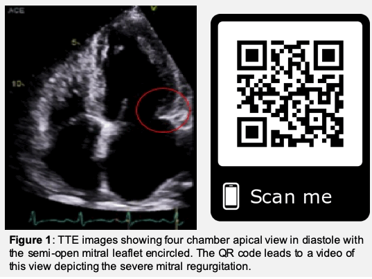
Case Presentation: Radiation associated cardiac disease (RACD) is an important adverse effect which may not present until years to decades following therapy. Presentations include arrhythmias, acute coronary syndromes, heart failure and valvular pathologies, as seen in this presentation. A 50-year-old female with past medical history of breast cancer treated with chemo-radiation eight years prior presented with respiratory distress. Transthoracic echocardiogram (TTE) revealed radiation-induced restricted motion of the posterior leaflet of the mitral valve in the semi-open position with severe mitral regurgitation (MR) and a posteriorly directed jet (Figure 1).
Discussion: Breast cancer is the most common malignancy in females with 1 in 8 US women developing invasive breast cancer [1]. Radiation associated cardiac disease (RACD) has been well studied in radiation of the mediastinum, chest wall and breast. Primary valvular pathology in RACD includes the mitral (43%) and aortic (37%) valves with pathophysiology being related to microvascular ischemic changes leading to fibrosis and calcification [2-3]. Most often, symptoms do not present until 4.5-16 years after initial treatments; however, increased radiation dose above 30 Gy may expedite RACD with findings within 1-2 years [4-5]
Conclusions: This case presents pulmonary edema secondary to MR directly related to radiotherapy, eight years prior. RACD is a known adverse effect in such patients and the pathophysiology must be understood by clinicians, particularly in this aging population with increasing cancer survivorship. Cardiovascular disease being the most important factor for mortality in cancer survivors, routine follow-ups with non-invasive cardiac imaging should be considered.