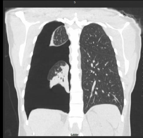
Case Presentation: 41 Years old Caucasian female with a past medical history of pleurisy since a year and iron deficiency anemia was present to the ED with three days history of right-sided chest pain and difficulty in breathing. – Vitals stable and chest x-ray, and CT of the chest showed sizeable right-sided pneumothorax got chest tube placement and repeat chest x-ray was showing re-inflation of lungs.
– But patient continued to have chest pain. Repeat CT chest which showed no improvement in the pneumothorax and trapped lung.
– Later patient underwent bronchoscopy which was normal, Video Assisted Thoracoscopy showed thick and erythematous plaques on pleura, and chronic adhesions of the right middle and lower lungs The patient underwent pleurodesis with the stripping of pleura and was discharged on contraceptive hormones .
– Biopsy was done , sent for cultures and immunostains which came back positive for endometriosis.
– Pt had a follow-up chest x-ray was improved with no evidence of pneumothorax .
Discussion: Catamenial pneumothorax is a manifestation of thoracic endometriosis in which the endometrium seeded thoracic cavity involving mostly the diaphragm ,pleura commonly presents as recurrent spontaneous pneumothorax up to 73%. Etiology has been remained undetermined and obscure. Most attributed theories were immunodeficiency, migratory or reflux theory and other proposed arguments are coelomic metaplasia and ectopic endometrial generation by stem cells and iatrogenic implantation. And proposed pathology was body responds to the proglandins released by the endometrial tissue especially constriction of bronchi and bronchioles and lead to bleb formation and further rutpure and spontanoeus pneumothorax.
Conclusions: Recurrent spontaneous pneumothorax in any menstruating female catamenial pneumothorax should be consideration .
As it should be managed surgically most of the times and medically with oral contraceptives for complete resolution.
Follow up with repeat chest x ray is warranted for further monitoring.