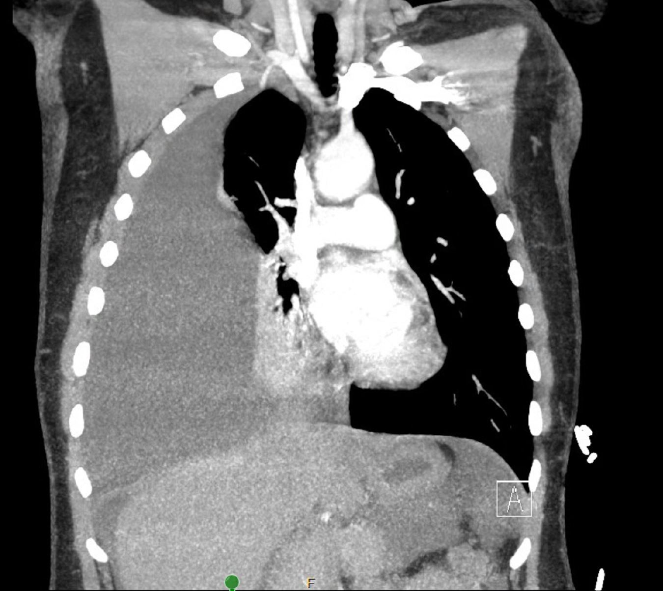
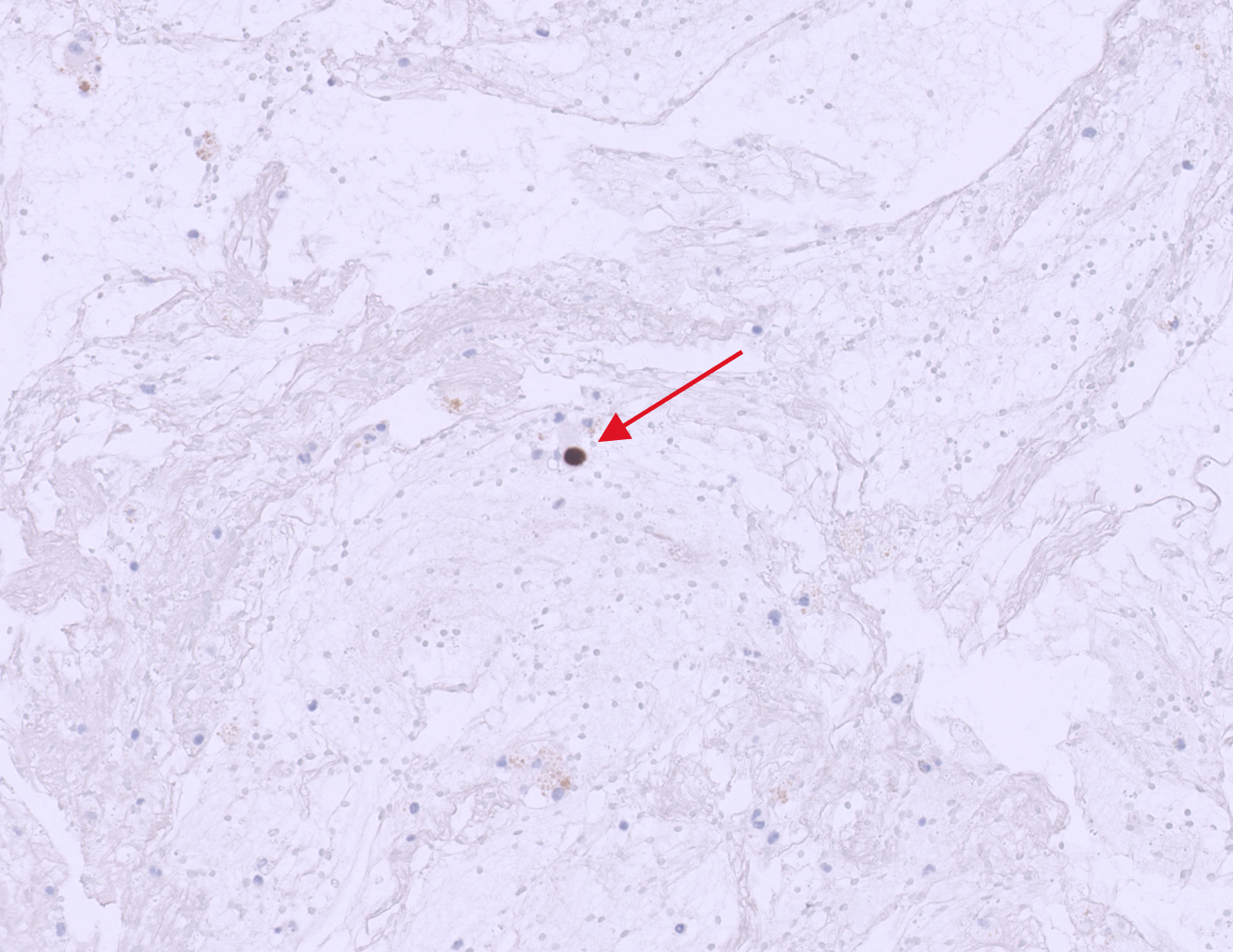
Case Presentation: A 38-year-old female with a past medical history of suspected endometriosis presented to the hospital with lightheadedness, palpitations, and chest tightness. Vital signs were remarkable for slightly elevated temperature (38.7 C) and tachycardia to 136 bpm. A contrasted chest CT demonstrated a large right-sided pleural effusion with complete atelectasis of right lower and middle lobes, with no evidence of pulmonary embolism (Figure 1). Pulmonology was consulted and performed a thoracentesis with 2200 ml of grossly brown fluid drained, and a right-sided pigtail chest tube was placed for further drainage. Post-procedure imaging demonstrated pneumothorax and thickened pleura, concerning for pneumothorax ex-vacuo due to a trapped lung. Thoracic surgery was consulted but deferred intervention for decortication given the patient’s clinical stability and minimal symptoms.Pleural fluid studies were consistent with exudative fluid and chronic hemothorax. There was no laboratory or clinical evidence of infection or autoimmune disease. Pleural fluid cytology was negative for malignant cells on two separate samples, and there was no evidence of malignancy on cross-sectional imaging, including pelvic MRI. Given the history of endometriosis, the pathologist performed PAX8 staining on pleural fluid, which was positive and highly suggestive of thoracic endometriosis (Figure 2). Gynecology was consulted and recommended initiating continuous oral contraceptives. The chest tube was removed, she remained stable and was able to be discharged home with close follow-up with pulmonology, gynecology, and thoracic surgery.
Discussion: This case demonstrates an unusual presentation of an already rare condition. The prevalence of thoracic endometriosis syndrome (TES) is estimated at 1.2 cases per 1,000 reproductive age women per year. TES most commonly presents as recurrent catamenial pneumothoraces. There have only been 176 cases of pleural effusion/chronic hemothorax secondary to TES identified and reported worldwide. The pathophysiology is not well-understood, but it is theorized to be due to migration of endometrial cells from the peritoneum to the pleura through defects in the diaphragm, hematogenous or lymphatic spread, or metaplastic transformation of pleural endothelial cells into endometrial like tissue. Among patients who undergo surgery, a diagnosis can be made when endometrial glands and stroma are present on a histologic sample. Immunohistochemical staining with ER, PR, CD10, IFITM1, and PAX8 can assist in making the diagnosis when histology is inconclusive or unavailable. PAX8 (Paired Box Gene 8) is a protein whose expression is almost exclusively found in the thyroid, kidney, and female reproductive tract organs. The positive PAX8 staining of the cells in the pleural fluid provided further support for the diagnosis of TES in our patient.
Conclusions: Hemorrhagic pleural effusion can be a rare manifestation of TES. Given its rarity, other more common causes must be thoroughly evaluated for and ruled out including malignancy, autoimmune disease, and infection. There are no standardized diagnostic criteria for TES. Recognition relies on multi-disciplinary evaluation and discussion amongst hospital providers and specialists including pulmonology, thoracic surgery, and gynecology.