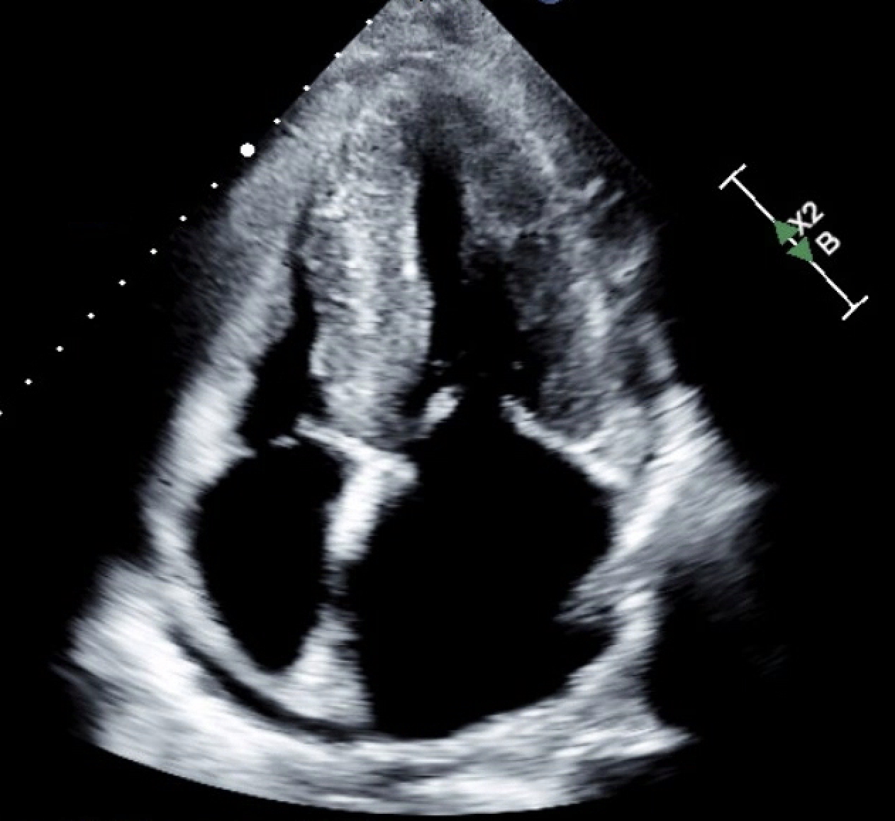
Case Presentation: We present a 65-year-old female patient with 3-4 months of dyspnea, initially treated outpatient for pneumonia without improvement. She presented to the Emergency Department (ED) with worsening symptoms, and physical examination revealed volume overload but no fever. Labs showed BNP elevated to 345 pg/mL and a normal white blood cell count. Computed Tomography (CT) angiography was negative for PE but demonstrated cardiomegaly, septal thickening, and bilateral pleural effusions, prompting admission for presumed heart failure exacerbation. Differential diagnosis included HFpEF, pulmonary hypertension, and arrhythmia. Infection, PE, and nephrotic syndrome were considered less likely. The patient received diuretics over the first 5 days with improvement in symptoms. Transthoracic echo (TTE) revealed an increased left ventricular wall thickness, global hypokinesis, ejection fraction of 45-50%, and diastolic dysfunction, concerning for an infiltrative myopathic process. Cardiovascular Magnetic Resonance (CMR) Imaging revealed septal hypertrophy and diffuse non-ischemic fibrosis. Guideline Directed Medical Therapy (GDMT) was not initiated due to concern for cardiac amyloidosis. SPEP, UPEP, and immunoglobulins showed mildly elevated serum kappa free light chains and remarkably elevated lambda free light chains (1048), consistent with AL amyloidosis. Hematology performed a bone marrow biopsy with myeloma panel. Further imaging ruled out multiple myeloma and transthyretin amyloidosis. The patient improved with diuresis and was discharged with close follow up with Hematology and Cardiology. Due to high suspicion for cardiac amyloid, home lisinopril and hydrochlorothiazide were discontinued, and spironolactone and low dose aspirin were started. She was referred for fat pad biopsy.
Discussion: While heart failure is common, cardiac amyloidosis is an uncommon but increasingly recognized cause of HFpEF(2,5). Its non-specific presentation is like other common causes of HFpEF, resulting in diagnostic delays. Imaging findings such as increased wall thickness are often attributed to hypertensive cardiomyopathy(3). However, timely diagnosis is critical as management and prognosis differ greatly from other types of heart failure. AL amyloidosis has a poor prognosis, with up to 43% of patients dying in the first year(5). Treatment also differs; while GDMT is standard for other forms of HFpEF, for cardiac amyloid, it can be poorly tolerated or even harmful due to preload dependence. Disease specific treatment can improve outcomes(1). This case underscores the need for high clinical suspicion when HFpEF is accompanied by atypical imaging findings. In this case, septal thickening, pleural effusions, and cardiomegaly raised the suspicion that this patient had more than simple heart failure.
Conclusions: Delayed diagnosis of cardiac amyloid is common. Clinicians should maintain a broad differential and high suspicion for uncommon causes of common conditions. This case illustrates the importance of avoiding anchoring bias, recognizing subtle imaging and laboratory evidence, and initiating timely diagnosis. Prompt identification and disease-specific therapy can improve outcomes.