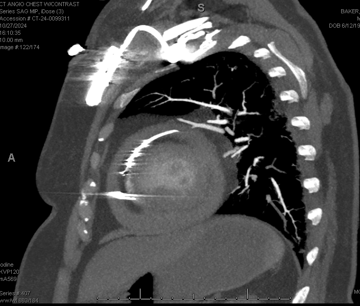
Case Presentation: A 63-year-old female with paroxysmal atrial fibrillation managed with apixaban status post biventricular pacemaker implantation a year ago presented with persistent chest pain and worsening dyspnea over the past week, accompanied by bilateral lower extremity pain. She reported a persistent “poking” sensation in her chest, particularly with movement. Vitals were stable and afebrile. On physical exam, patient had significant mottling of the lower extremities. Laboratory examination was significant for elevated leukocyte count. CT scan of the chest with contract revealed perforation of the right ventricular wall by the pacemaker lead, with a moderate pericardial effusion without tamponade. Patient was admitted to the intensive care unit for monitoring and potential surgical intervention. A decision was made to proceed with thoracotomy to extract and replace the RV lead and repair the myocardial wall. Inspection revealed that a ventricular lead had perforated through the apical region of the right ventricle adjacent to the septum. Surgery was successful, and postoperative echocardiography confirmed a stable pericardial effusion without progression.
Discussion: Pacemaker lead perforation is a rare but critical complication, with a general prevalence of 0.1-6%. Chronic perforation, defined as occurring more than 30 days post-implantation, is even rarer, affecting less than 1% of patients. We describe a rare instance of lead perforation into the right ventricle, resulting in significant pericardial effusion. It is crucial to maintain vigilance in pacemaker-dependent patients presenting with chest pain, as even though perforation is rare, it is a life-threatening complication that can present in a variety of ways, from stable cases with chest pain, as in our patient, to hemodynamically unstable cases with tamponade.
Conclusions: Chronic right ventricular lead perforation, though uncommon, requires rapid identification and a thorough risk assessment. Through this case, we emphasize the need for heightened clinical awareness in the management of patients with implanted cardiac devices.