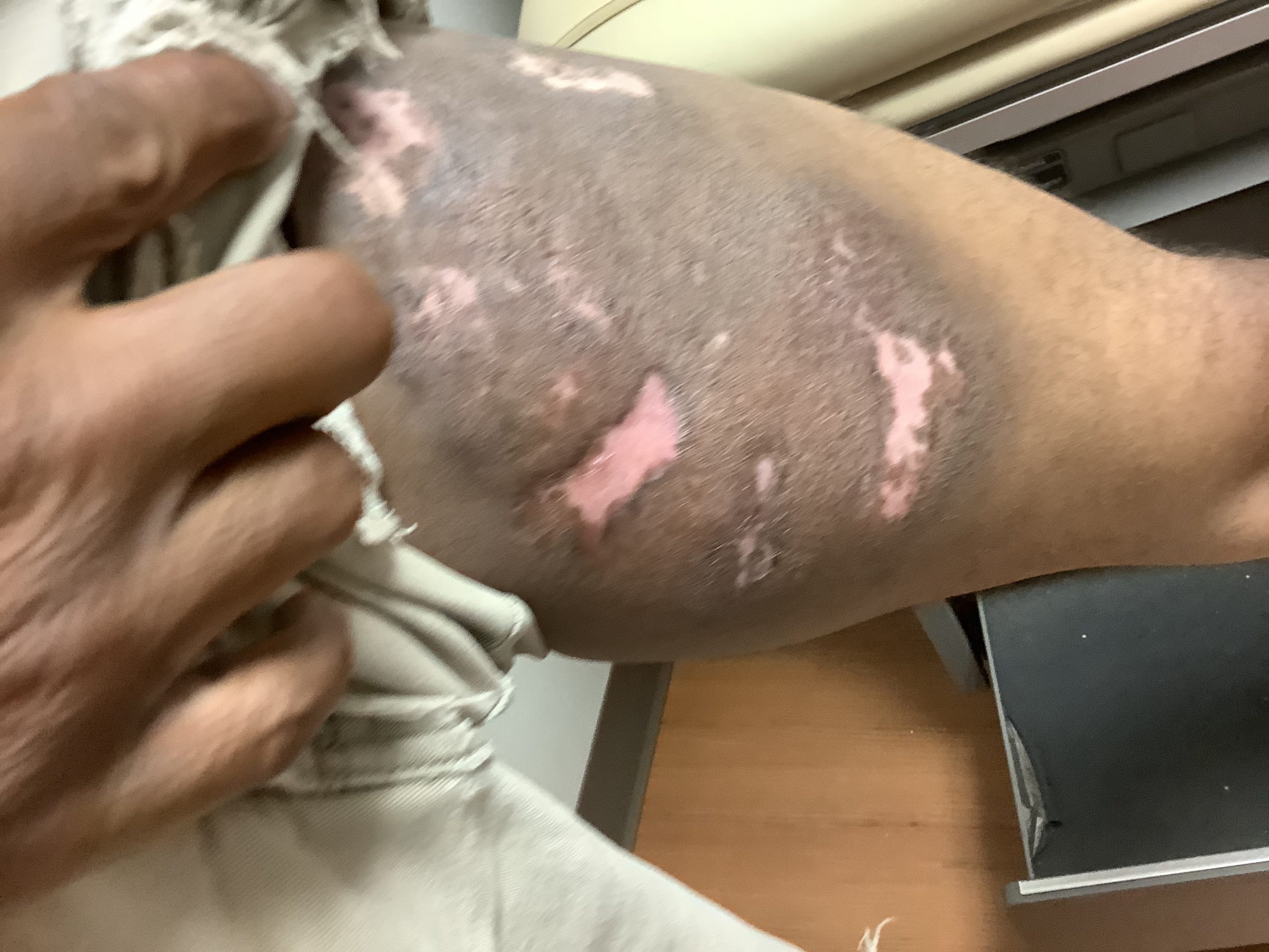
Case Presentation: A 45-year-old African American male without significant past medical history presented with a worsening left groin wound. He reported a slow growing, left thigh mass present for the past two years. Exam findings showed multiple areas of necrotic tissue with ulcerated lesions containing foul smelling purulent drainage, with associated left inguinal lymphadenopathy. CT scan of left lower extremity demonstrated a fungating mass extending into the deep tissues with no intramuscular infiltration. Skin biopsy results confirmed T cell lymphoproliferative disorder with CD 3/4 positive, CD 30 negative with TCR rearrangement. Six-month follow up confirmed resolution of T cell lymphoma following surgical excision and 15 cycles of radiation.
Discussion: Cutaneous T cell lymphoma can present as thin plaques and progress to violaceous ulcerated lesions with localized lymph node involvement. Diagnosis is based on skin biopsy with proper immunohistochemistry and molecular analysis. T cell gene rearrangement analysis can help detect CTCL in early stages. Imaging of surrounding tissues via CT can help detect underlying lymph node and solid organ involvement. Radiation therapy is used for localized disease; however, there is lifetime risk of recurrence. Other options of treatment for localized disease include topical chemotherapy agents. If CTCL becomes refractory to localized treatment or has spread to solid organs, high dose of multi-agent systemic chemotherapy is warranted.
Conclusions: Any slow growing fungating skin lesion should undergo a skin biopsy to assess for malignancy despite presentation of suspected infectious process. Early detection of Cutaneous T cell lymphoma would lead to localized treatment with radiation and allow for overall good prognosis without the need for systemic chemotherapy.