Case Presentation: A 62-year-old woman presented with a three week history of worsening shortness of breath. She endorsed a dry non-productive cough of several days without fever, chills, or other symptoms. Review of systems was negative for hemoptysis. Past medical history includes HFpEF, hypertension, and chronic atrial fibrillation on apixiban.
On presentation, her temperature was 36.3 C, HR 50bpm, BP 194/65 mmHg, RR 24 bpm, and O2 saturation 92% on room air. Physical exam revealed a woman in mild respiratory distress. She was bradycardic with an irregularly, irregular rhythm. Jugular venous distension was not present. Crackles were present on auscultation. Extremities revealed edema. Labs revealed a WBC of 12.3k/mm3 and BNP 186. Chest radiograph was consistent with pulmonary edema.
The initial diagnosis was acute decompensated heart failure. After diuresis, her shortness of breath did not improve. Although antibiotics were started for a potential infectious etiology, she did not improve. A ventilation/perfusion scan returned low probability for pulmonary embolism. Synchronized cardioversion from atrial fibrillation to sinus rhythm did not improve her symptoms.
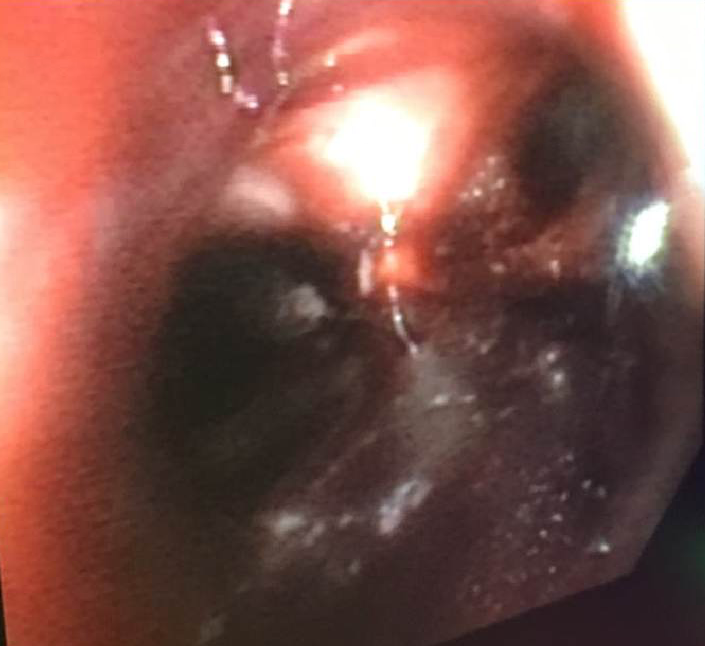
The patient’s hemoglobin dropped from 8.1 g/dL on admission to 6.4 g/dL and her oxygen requirements increased. She was eventually placed on high flow nasal cannula and developed acute hypoxemic respiratory failure requiring intubation and mechanical ventilation. Bronchoscopy visualized diffuse frank blood in both lungs consistent with diffuse alveolar hemorrhage. Anticoagulation was discontinued, and she was transfused packed red blood cells. Viral panel from the bronchoscopy was positive for rhinovirus. For the presumed diagnosis of diffuse alveolar hemorrhage, she was started on high dose steroids and continued on mechanical ventilation for airway protection.
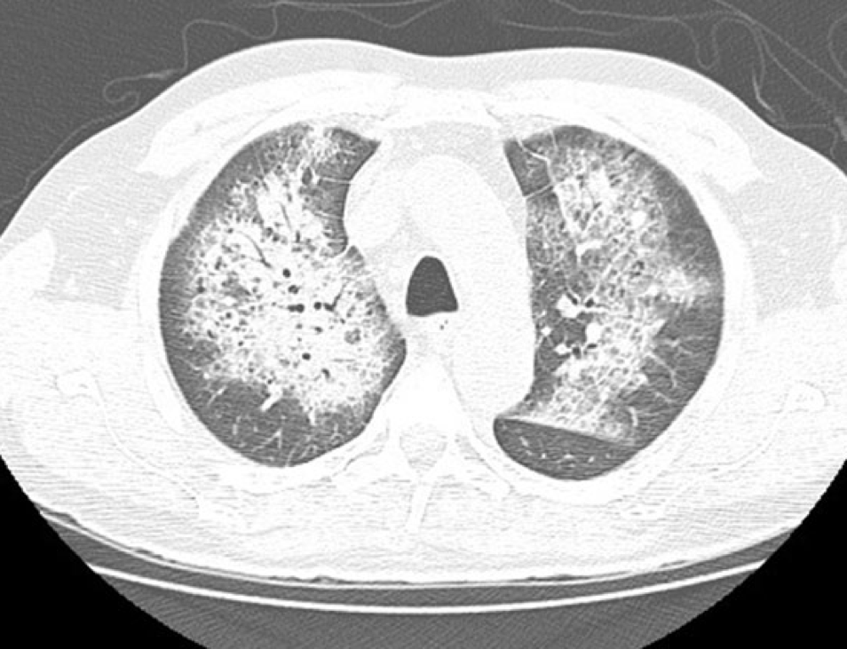
Discussion: Acute hypoxemic respiratory failure is a common diagnosis encountered by hospitalists. Diffuse alveolar hemorrhage (DAH), although an infrequent cause of acute hypoxemic respiratory failure, cannot be missed. Typical features of DAH include hemoptysis with diffuse alveolar infiltrates on imaging. Interestingly, our patient did not experience hemoptysis.
Bronchoscopy is warranted in patients with acute hypoxemic respiratory failure of unknown etiology, as it can help identify uncommon conditions such as DAH. The most likely trigger for DAH in this patient was superficial vessel irritation from rhinovirus pneumonia in the setting of anticoagulation. Acute hypoxemic respiratory failure of unknown etiology should prompt urgent consideration of bronchoscopy.
DAH is most commonly caused by vasculitic and autoimmune disorders; as such, steroids should be initiated during the diagnostic workup. Less common etiologies include coagulation disorders, infection, and drug toxicities. DAH should be considered as an uncommon yet very important etiology of acute hypoxemic respiratory failure, even in the absence of hemoptysis.
Conclusions: Physicians must recognize that an atypical presentation for acute hypoxemic respiratory failure could be secondary to Diffuse Alveolar Hemorrhage. Typical features of DAH include respiratory compromise and hemoptysis with progressive alveolar infiltrates on chest imaging. Although DAH is most commonly caused by vasculitic and autoimmune disorders, it can occur secondary to medication side effects. Bronchoscopy is an important diagnostic modality in the setting of acute hypoxemic respiratory failure of unknown etiology.