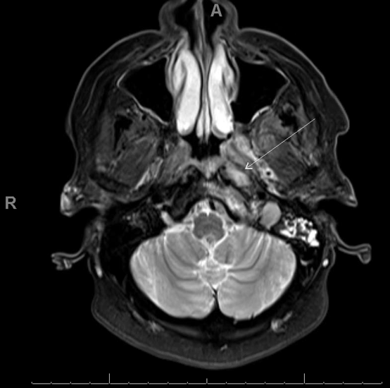
Case Presentation: A 64-year-old woman with uncontrolled type 2 diabetes mellitus presented with five months of constant, stabbing left temporal headache radiating to the ear, jaw, and neck. She had associated progressive neurologic deficits, including blurred vision, flashing lights, and subacute left-sided hearing loss. She had presented to another hospital two months prior to the current presentation and was diagnosed with giant cell arteritis despite negative bilateral temporal artery biopsy. She was treated empirically with high-dose steroids, but her symptoms continued to worsen. On presentation, she was afebrile with normal visual acuity with corrective lenses, diminished left-sided hearing, and mild tenderness to palpation of the left temple, ear, and jaw. Labs revealed glycated hemoglobin exceeding 14% and elevated inflammatory markers with ESR of 111 mm/hr and CRP of 22 mg/L. Computed tomography of the head demonstrated a left retropharyngeal soft tissue lesion with bony erosion of the left lateral clivus and sphenoid sinus opacification. Brain magnetic resonance imaging (MRI) showed an infiltrative enhancing soft tissue lesion centered at the left cranio-cervical junction with bony involvement suggestive of an inflammatory process. The patient underwent left concha bullectomy and left sphenoidotomy with debridement. Intra-operative cultures were positive for Staphylococcus aureus and Klebsiella pneumoniae, while surgical histopathology showed chronic sino-nasal mucosal inflammation with fungal balls. The patient was treated with broad-spectrum antibiotics and antifungals for polymicrobial skull base osteomyelitis. Her headache and neurologic symptoms completely resolved after six weeks of treatment.
Discussion: Skull base osteomyelitis is a rare and potentially fatal clinical entity caused by invasive fungal or bacterial infection from a sinus or otologic source. Uncontrolled diabetes, as in this patient, is a leading risk factor due to the associated immunosuppression and microangiopathy. Depending on the affected skull bones, symptoms are often vague and non-specific with cranial neuropathies occurring late in disease. MRI is the preferred imaging modality, but biopsy is typically required for definitive diagnosis and to rule out alternative diagnoses. Even then, identification of the causative microorganism can be complicated by false-negative results, particularly in the clivus bone and preclival soft tissues where chronic inflammation can be low-grade and pathogen burden low. Prolonged antimicrobial treatment is the cornerstone of treatment, often with surgical debridement. Timely intervention is needed to prevent the sequelae of untreated disease, namely neuro-invasive disease, such as meningeal or parenchymal involvement. The proper diagnosis and management of skull base osteomyelitis depends on a high degree of clinical suspicion and a well-executed, multidisciplinary approach.
Conclusions: Skull base osteomyelitis is a rare clinical entity typically seen in elderly diabetic and immunocompromised patients. Its presentation can mimic other non-infectious inflammatory and malignant processes so is often misdiagnosed. Clinicians must maintain a high index of suspicion until a definitive diagnosis is made, as to allow for appropriate intervention and prevention of associated complications.