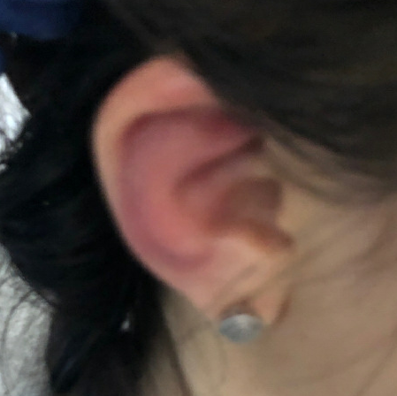
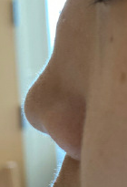
Case Presentation: A 20-year-old female with a history of mild intermittent asthma and seasonal allergies presented to the Emergency Department with two months of pleuritic chest pain, fevers, weight loss, and hoarseness along with one day of right ear erythema and swelling. Her symptoms had improved with steroids but returned with weaning. A previous extensive work-up including bone marrow biopsy, CT scan, echocardiogram, tagged WBC scan, and infectious plus autoimmune work-up failed to reveal a diagnosis. Key exam findings on admission included temperature 38.4 degrees C, bilateral erythematous pinnae with lobule-sparing, slight depression in nasal cartilage, dysphonia, and tachypnea with splinting. Laboratory studies revealed an ESR of 129 mm and CRP of 27.6 mg/dL. A flexible laryngoscopy demonstrated hemorrhagic edema of the left true vocal cord. A bronchoscopy was performed with findings of severe tracheobronchomalacia and edematous airways. Right helix biopsy demonstrated cartilaginous focal loss of nuclei and necrosis consistent with the diagnosis of relapsing polychondritis with diagnosis further supported by elevated serum collagen type II antibodies. The patient was started on IV methylprednisolone, methotrexate, and infliximab with significant improvement in all symptoms.
Discussion: Relapsing polychondritis is a rare disease with many non-specific symptoms that can mimic many other inflammatory or hematological conditions. Auricular involvement such as chondritis was the key pivot point in diagnosis in this case and overall is the most common feature of relapsing polychondritis. It occurs in 90% of patients but is only present in 40% on initial presentation. As seen in this case, other common manifestations of cartilaginous inflammation include costochondritis, nasal deformities, and large airway involvement with the latter potentially leading to life threatening disease. Treatment options vary depending on severity of disease and if associated with coexistent disease.
Conclusions: This case highlights the difficulty in establishing a rare diagnosis with several non-specific features, the importance of a single physical exam finding to alter diagnostic work-up, and importance of early recognition given risk of severe airway manifestations.