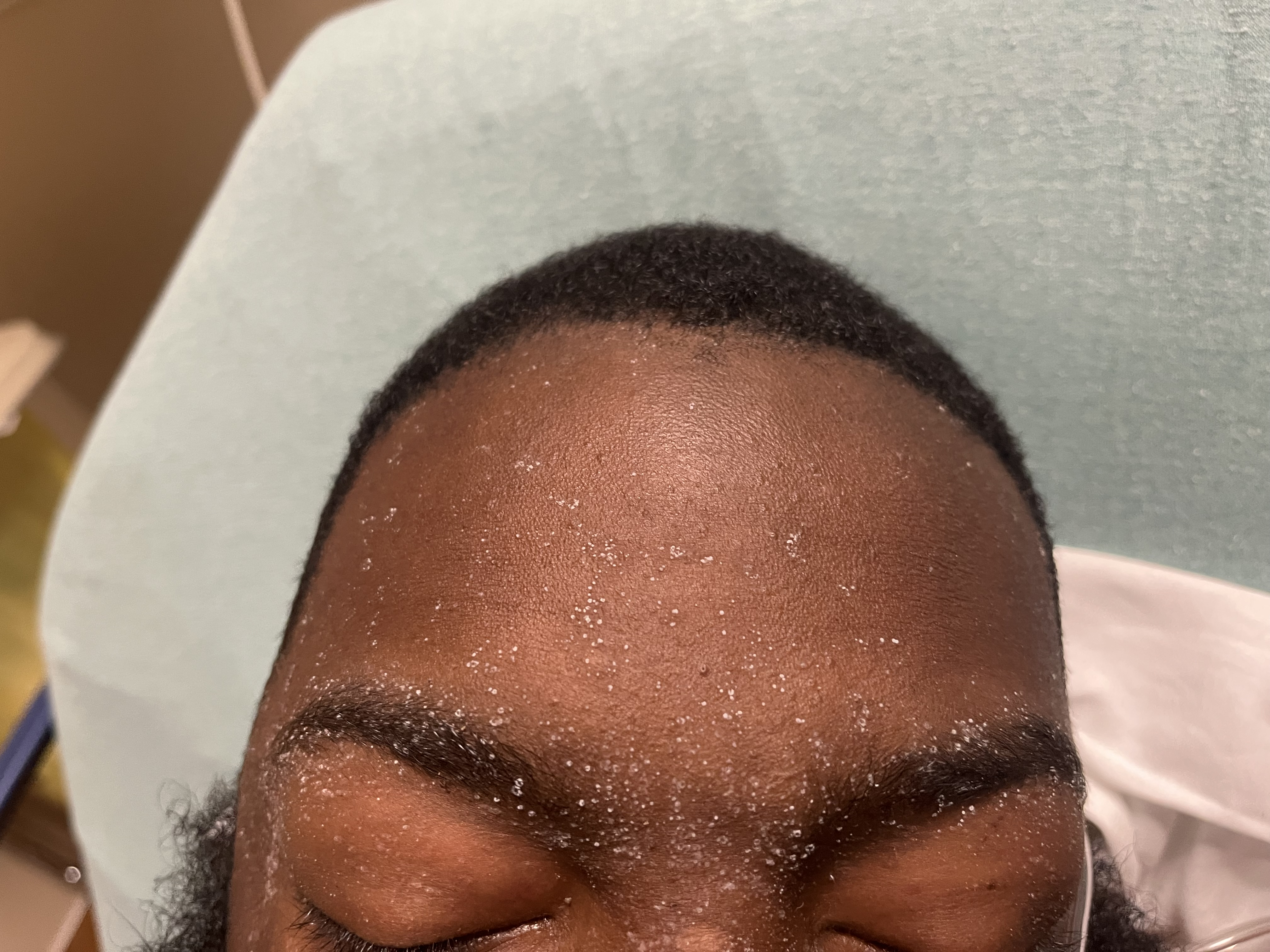
Case Presentation: A 34-year-old African American male with history of HTN, HLD, obesity, T2DM complicated with ESRD on hemodialysis the past 2 years presented to our EC in Houston for progressively worsening dyspnea, volume overload and confusion. The patient had been previously receiving regular hemodialysis three times weekly. However, due to lack of transportation, he had been unable to get to dialysis for five months prior to admission. He had also been unable to afford medication for over a year. On exam in the EC, the patient was lethargic, hypertensive, hypoxic, with a BMI 50.2 and he had edema all the way to his chest with peau d’ orange appearance. Prominently, he had white crystals (size ~0.5 mm) over his forehead, bilateral eyebrows, and over the bridge of his nose. Patient was found to have Hgb of 5 g/dL, K of 5.9 meq/L, BUN 168 mg/dL, Cr 21.1 mg/dL, phosphorus 10.9 mg/dL, HgbA1c 5.0 %. Patient was admitted and emergently dialyzed. Over a course of 3 hemodialysis sessions in 5 days, the patient’s BUN went down from 16813210345 mg/dL. His mental status also improved. His forehead crystals resolved. He was reassigned to a new dialysis facility after social issues were resolved and discharged home after prolonged 15-day hospital stay.
Discussion: Uremic frost, first described in 1865 by Hirschsprung, was seen commonly in end stage renal disease (ESRD) patients until the advent of hemodialysis since 1950s. It is now a rarity in developed countries with hemodialysis widely available. The pathophysiology is postulated that high concentrations of urea in the serum allows for urea accumulation in the dermis, where it leeches into sweat glands and surfaced on skin. Drying through evaporation yields white or yellow colored uremic frost. The exact mechanism has never been studied in vitro. Uremic frost has a predilection of skin areas associated with higher pilosebaceous unit distribution in the face, neck, scalp, forearms and chest. Usually, it is associated with a BUN level of 71 mmol/L (200 mg/dL). In the emergent settings, especially resource scarce parts of the world or inadequate lab access, this specific dermatological sign helps clinicians identify patients with severe underlying renal diseases. Definitive diagnosis requires scraping of crystals for urea content analysis. The differential diagnosis of uremic frost includes, atopic dermatitis, retention keratosis, and post inflammatory desquamation. In patients with cystic fibrosis, crystal analysis is important, as sodium chloride crystal formation on skin has been reported. Gout and oxalosis can also present as skin crystals but usually intra-dermis and not on the surface of skin. The treatment of uremic frost is dependent on the correction of underlying renal dysfunction, and the skin findings completely (or usually completely) resolve with dialysis.
Conclusions: Once a common presentation of end stage renal failure now becoming rare in developed countries, uremic frost is still a valuable clinical exam finding in resource scarce areas of the world as it suggests an emergent indication for dialysis. Complete resolution of uremic frost can be achieved with adequate hemodialysis


