Case Presentation: A 33-year-old woman with past medical history of hypertension presented to an outside hospital with a one week history of dyspnea on exertion and acute hypoxic respiratory failure. She had also previously lived in Arizona.
Upon presentation to outside emergency department, she had significant sinus tachycardia and tachypnea with an oxygen saturation of 68% on room air. Her systolic blood pressures were consistently greater than 200. She was then intubated, but subsequently went into pulseless electrical activity. She was resuscitated successfully. Chest X-ray revealed diffuse opacities consistent with possibly ARDS.
Upon arrival, she was sedated and intubated with a blood pressure of 155/105 mmHg. Exam and bedside ultrasound suggested pulmonary edema, and her lower extremities were warm and pedal pulses were palpable at that time.
She became hyperkalemic and anuric and was started on Intermittent hemodialysis. Her blood pressures were slower lowered, and she then developed pulseless lower extremities.
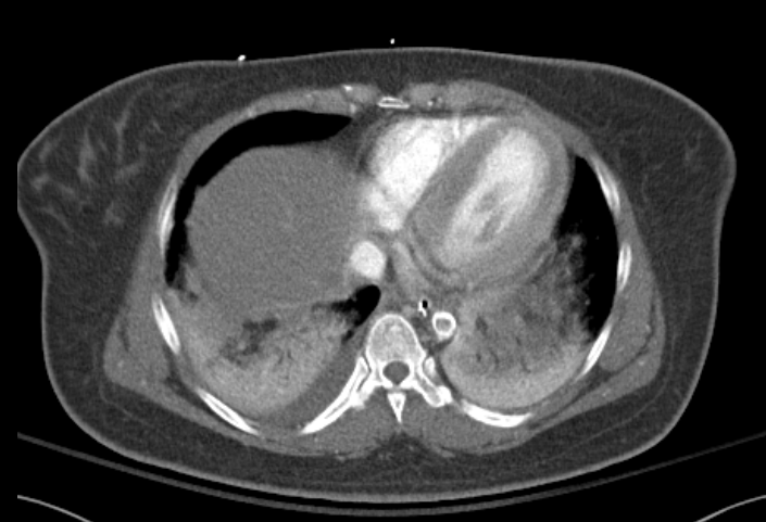
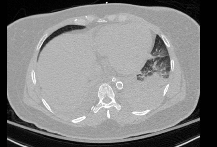
CT chest abdomen pelvis showed a calcified thrombus in the suprarenal aorta consistent with coral reef aorta.
She then underwent a vascular surgery operation to bypass the aortic occlusion from the common femoral artery which allowed retrograde revascularization of the kidneys. She was eventually discharged home, regained renal function, and was able to return to her medical baseline.
Discussion: Coral reef aorta is a rare vascular disease of the that is characterized by rough, calcified plaques that extend into the lumen resulting in decreased vascular flow. These plaques are often described similar to bony lesions because of their high percentage of calcium
The clinical presentation of coral reef aorta depends on the vessels involved. Similar to other causes of aortic coarctation, patients can have intermittent claudication in the lower extremities. Physical examination often reveals diminished lower extremities pulses and arterial perfusion.
Management is usually surgical thromboendarterectomy or a bypass graft.
She was initially thought to have ARDS secondary pulmonary infection, but CT showing the coarctation likley led to flash pulmonary edema secondary to hypertension due to the occlusion. Her renal failure was also secondary to hypoperfusion.
Conclusions: Coral reef aorta should be in the differential diagnosis for patients with pulmonary edema and symptoms of aortic coarctation. Additionally, the mean arterial pressure goals may need to be considerably higher than expected because of the amount of pressure required to avoid hypoperfusing vasculature downstream of the occlusion.