Case Presentation: A previously healthy 11-month-old female was admitted to the hospital with new onset of skin lesions. One month prior to admission, her parents noticed “bumps on her bottom.” They initially thought the bumps were secondary to insect bites, and took her to a local urgent care, where she was prescribed topical antibiotic ointment. The lesions persisted, and she developed new papules on her arms and legs, and fever to 101°F. At this time, her pediatrician evaluated her and diagnosed hand, foot, and mouth disease. She defervesced the next day, but the lesions persisted with development of purulent drainage, crusting, and eventual ulceration and pain. Repeat evaluation resulted in prescription of cephalexin for presumed impetigo. With failure to improve, she also completed a course of sulfamethoxazole/trimethoprim as well as topical and oral acyclovir without improvement. Bacterial and viral cultures of the lesions were negative. Given persistent pain and severity of the lesions, she was admitted to the hospital.
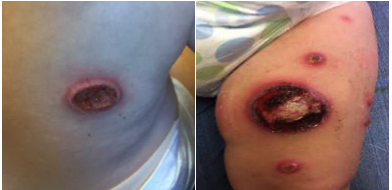
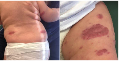
On admission, she appeared non-toxic but in significant pain. Her exam revealed diffuse well-circumscribed erythematous papules and pustules of varying sizes on her face, legs, arms, hands, and feet intermixed with well circumscribed erosions with central crusting and a violaceous border (Figure 1). Initial differential diagnosis included atypical mycobacteria infection, nodular vasculitis, T-cell cutaneous lymphoma, and infantile pyoderma gangrenosum. Labs were significant for a leukocytosis with neutrophilia; LDH and uric acid were within normal limits. Intravenous antimicrobials were started on admission but discontinued when repeat cultures were negative. Dermatology performed a skin biopsy which was consistent with pyoderma gangrenosum. Oral steroids were started with overall improvement and resolution of several lesions. She was discharged home on oral steroids.
Two weeks after discharge, she experienced worsening pain and new skin lesions. She was admitted to the hospital for IV steroids. She was later started on immunomodulatory therapy with Infliximab after discussion with rheumatology and dermatology. Given potential for associated diseases, she was evaluated for underlying systemic disorders with normal immunoglobulin levels, blood smear, diphtheria and tetanus antibody titers, leukocyte adhesion deficiency panel, and neutrophil function test. At this time, no other comorbid conditions have been diagnosed in this patient. She was discharged home on oral steroids and the plan for monthly Infliximab infusions with rheumatology and dermatology follow up.
Discussion: Infantile pyoderma gangrenosum (IPG) is very rare with less than 20 cases reported in the literature. It is a neutrophilic dermatosis that presents as ulcerations of the skin with inflammation. Infants diagnosed with IPG warrant further work-up for associated systemic diseases including inflammatory bowel disease, Takayasu’s arteritis, leukocyte adhesion deficiency, and chronic granulomatous disease. Most cases, however, are idiopathic—though it can be difficult to rule out an associated condition, given skin findings may precede other systemic symptoms by several years. At this time, standardizing treatment is challenging given the rarity of IPG, but includes immunomodulation with steroids and/or biologic agents.
Conclusions: Though rare in infants, pyoderma gangrenosum should be considered in the differential diagnosis for an infant with diffuse ulcerative skin lesions.