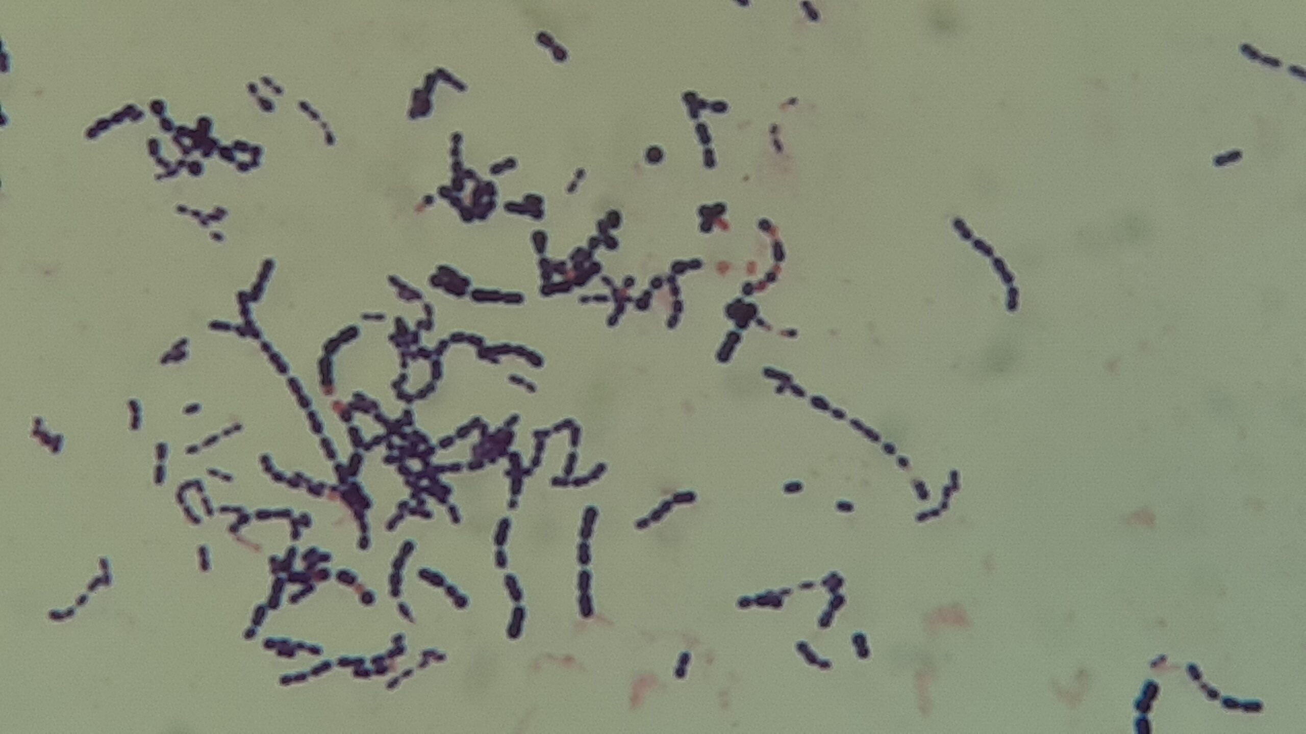
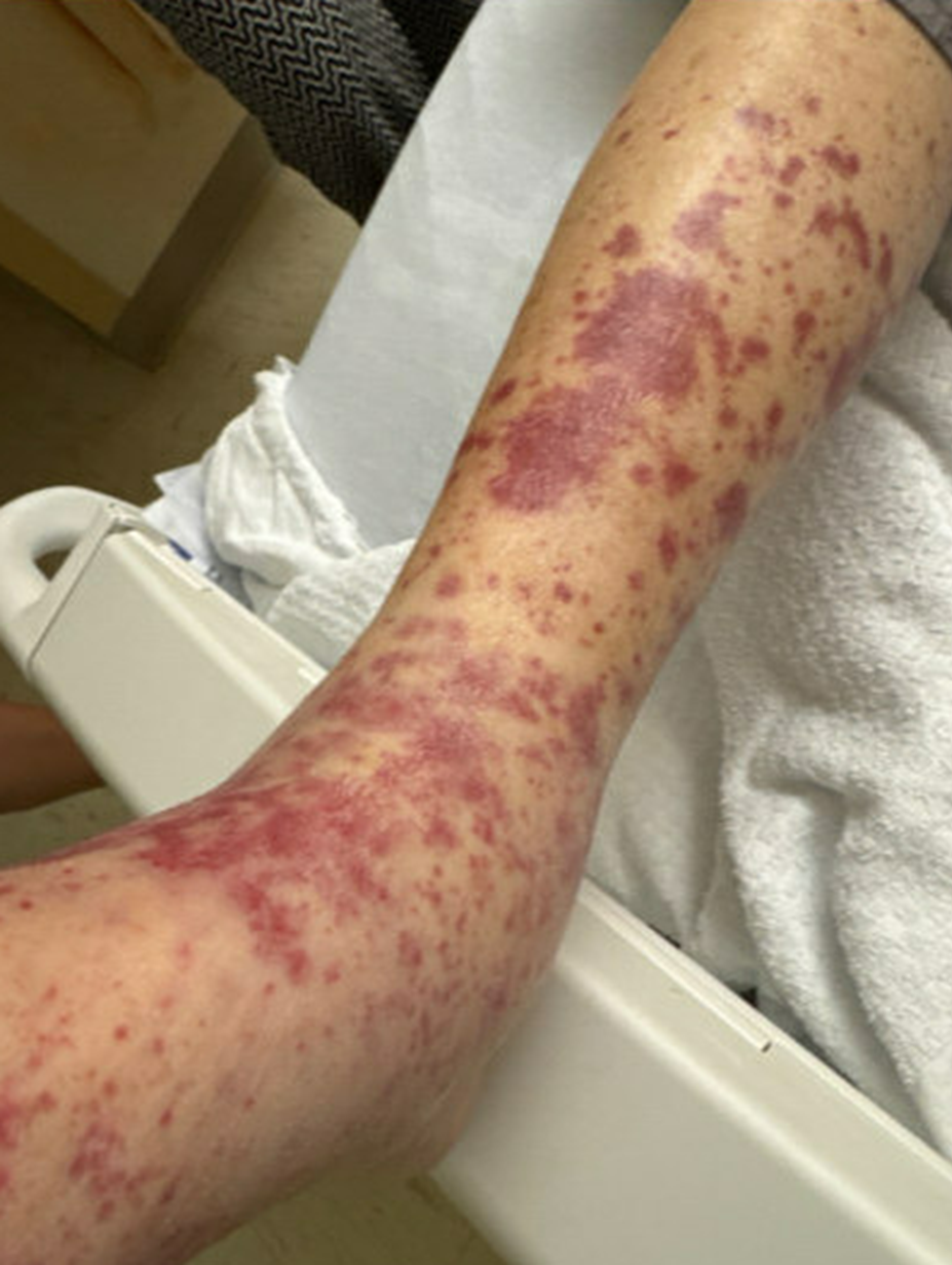
Case Presentation: A 74-year male with a medical history of CAD, aortic stenosis (s/p TAVR), type 2 diabetes, andHTN, presented to the emergency department with a chief complaint of shortness of breath and darkurine. 11 months prior to admission, the patient underwent a routine dental procedure. 10 months prior tohospitalization, he had been experiencing progressive dyspnea and fatigue with a nonproductive cough. 3months prior to hospitalization, he noted a 20lb weight loss, weakness, and a rash of the lower extremitiesthat had been waxing and waning. He also noted prior to hospitalization dark, “Coke colored” urine. Inthe time leading up to admission, he had undergone an evaluation for anemia and hematuria, which wereunrevealing. On admission to the hospital, physical exam was remarkable for trace peripheral edema,systolic murmur, and petechial rash in the lower extremities. Laboratory findings included hemoglobin of8.1 g/dL, WBC count of 12.8 ×10³/, elevated creatinine of 1.5 mg/dL, elevated Pro-BNP of 5170 pg/mL.Urinalysis showed large blood with 51–100 RBC per HPF. Additional labs included a CRP of 13.0 mg/dLand ESR of 101 mm/h, low complement levels with a C3 52mg/dL and C4 <8mg/dL. ANCA IgG andanti-GBM were negative. A chest x-ray was suggestive of mild pulmonary edema. Shortly after admission, blood cultures returned positive for gram-positive cocci in pairs andchains, prompting the initiation of ceftriaxone. Transthoracic echocardiogram was notable for normal EFwith mild to moderate mitral regurgitation. A punch biopsy of his purpuric rash revealed leukocytoclasticvasculitis. A transesophageal echocardiogram showed thickening of the posterior leaflet tip of the mitralvalve—findings congruent with a vegetation. Blood cultures speciated to Gemella but sensitivities couldnot be performed due to poor organism growth. Repeat blood cultures were negative. The patient wasdischarged on cefdinir for 4 weeks with dalbavancin for 6 weeks and in 3-4 weeks post hospitalization, hereported a resolution of all symptoms.
Discussion: Subacute infective endocarditis remains a challenging diagnosis due to its variable clinicalmanifestations. While Staphylococcus and Streptococcus species account for many cases of endocarditis,uncommon pathogens such as the Gemella species have increasingly been recognized. Gemella are gram-positive cocci that are part of the normal oropharyngeal and gastrointestinal flora but rarely can causesystemic infections. Gemella infective endocarditis accounts for approximately 0.4% of reported cases. Inaddition, Gemella has shown poor growth on blood cultures through typical methods in hospitallaboratories.In addition to valvular complications, manifestations of infective endocarditis include immune-mediated processes such as immunocomplex glomerulonephritis and, rarely, leukocytoclastic vasculitisseen in our case. In our patient, the Gemella species infective endocarditis led to immune complexmediated disease, as evidenced by the low circulating complement levels.
Conclusions: The case presented demonstrates a rare instance of Gemella subacute infectious carditis withsequelae of immune complex deposition. The constellation of hypocomplementemia, vasculitic eruptions,and unexplained hematuria in the setting of systemic symptoms, along with known difficultly growingatypical organisms on blood cultures such as Gemella, should alert the hospitalist to evaluate for thepresence of infective endocarditis.