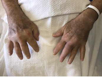
Case Presentation: A 73-year-old Iraqi man with asthma and type 2 diabetes who enjoys gardening was diagnosed with seronegative rheumatoid arthritis (RA) when he presented with joint pain and swelling in both hands and wrists. He was treated with prednisone (20 mg daily) and methotrexate. One month later, he developed swelling and ulcerative papules on the left leg. Over the next six months, he had no improvement in joint pain and the skin lesions had progressed to also involve both arms. Skin biopsies from the lesions of his left arm and leg were positive for acid-fast bacilli (AFB), for which he was admitted to an outside hospital for possible disseminated tuberculosis. Cultures eventually grew Mycobacterium chelonae. Treatment was switched from prednisone and methotrexate to hydroxychloroquine and he was discharged without antibiotics. He presented to our hospital for a second opinion. He reported an unintentional weight loss of 16 lbs. over the last month, fatigue, dyspnea, and 10/10 pain in both hands (figure) and knees. He was afebrile. Chest auscultation revealed global wheezing. A CT chest showed ground-glass opacities and radiographic studies showed findings suggestive of osteomyelitis and tenosynovitis in both hands. The diagnosis of seronegative RA was refuted, and he was admitted for probable disseminated nontuberculous mycobacterial (NTM) disease. He underwent surgical washout of the left hand for purulent septic arthritis. Synovial and bone biopsies showed granulomatous inflammation with AFB. He was empirically treated with tobramycin, clarithromycin, and levofloxacin for presumed M. chelonae. Levofloxacin was later changed to imipenem based on culture sensitivities.
Discussion: M. chelonae is a NTM generally found in water and soil, most commonly reported to cause skin and soft tissue infections. German researcher Friedrich Freidmann first isolated this pathogen from the lungs of sea turtles (Chelona corticata) in 1903, referring to it as the “turtle tubercle bacillus” (1). NTM infections can be tricky to diagnose because of their indolent course resulting in treatment delays. In this case, our patient presented with symmetric polyarthritis. Without classic RA x-ray findings and negative lab markers, he was diagnosed and treated for seronegative arthritis. Without fever or other systemic symptoms, infectious causes were low on the initial differential. However, after his lack of response to appropriate RA therapy and new skin findings, the differential diagnosis was not revisited. His infection blossomed on immunosuppressants with rapid progression and involvement of the skin, joints and lungs. M. chelonae can cause osteomyelitis in immunosuppressed patients although lung involvement is rare (2). Because of the extent of his infection, our patient required washouts of the infected joints and will remain on antibiotic therapy for several months paired with rehabilitation to improve function.
Conclusions: A broad differential diagnosis should be considered in evaluating patients with polyarthritis, including inflammatory disorders, infectious, neoplastic and drug-related causes (3). NTM infections such as M. chelonae, although rare, can present with extrapulmonary manifestations. Given that we generally view the timeline for an infectious etiology to be relatively short, we should keep this indolent organism that has a long incubation period in mind when evaluating patients with subacute or chronic joint and skin complaints that do not clearly fit into other illness scripts.