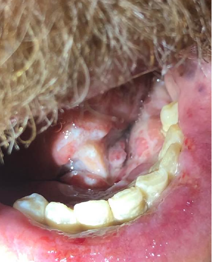
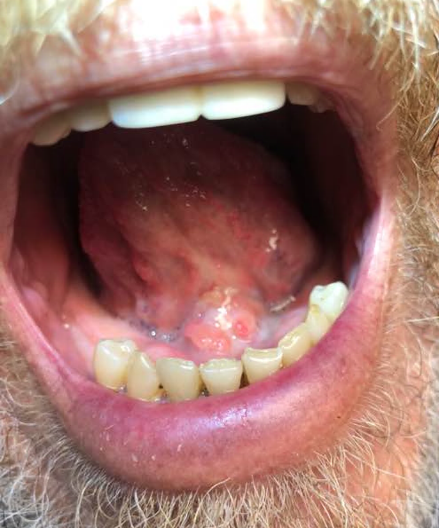
Case Presentation: A 63-year-old man was admitted to the hospital medicine service at a quaternary center for painful oral lesions that appeared 6 months prior. The lesions had started as multifocal soreness in the mouth floor 2 weeks after having undergone dental work. He was then evaluated by local oral surgery team who performed biopsies of the foci of abnormal mucosa and the patient was told no malignancy was identified. The sore areas later evolved into ulcers and the patient sought medical attention from a local rheumatologist. A second biopsy was done and was interpreted as oral chronic granulomatous disease. The patient was subsequently started on Prednisone and Clindamycin without significant improvement. The lesions then started to progress and extend beyond the floor of the mouth to involve the lower lip and tongue. They became very painful to the extent that the patient was unable to tolerate oral intake and feeding via percutaneous gastrostomy tube was being considered. He was unable to talk and communication with him was via writing. At that point, he decided to present to our center’s emergency department. There were no other symptoms. Medical history was remarkable for coronary artery disease, osteoarthritis, and hypertension. Social history was notable for tobacco use. There was no prior travel outside Tennessee. He was not on any medication known to cause mucosal ulcerations. On exam, his vital signs were normal and oral exam revealed multiple erythematous, tender ulcerated oral lesions (picture 1) with leukoplakia. Laboratory testing including that of HIV, herpes virus, vasculitis, and blood culture was unrevealing. Histoplasma antigen was positive. Chest CT did not show evidence of histoplasmosis. Upper and lower endoscopy evaluating for Crohn’s disease, given the granulomatous nature of oral lesions, were unrevealing. After multidisciplinary discussion with consultants from Rheumatology, Gastroenterology, Infectious disease (ID), and Oral surgery, the decision was to re-biopsy the lesions. Fungal culture of the specimen showed Histoplasma Capsulatum. Within 48 hours of initiation of Itraconazole, the patient started to improve and tolerate soft diet. Two weeks later, he was seen in the ID clinic and remarkable symptomatic improvement was reported by the patient. He was eating and talking normally. Exam showed residual oral edema and erythema without ulceration (picture 2).
Discussion: Histoplasmosis is a systemic granulomatous fungal infection caused by Histoplasma Capsulatum. Its clinical presentation was first described by Samuel Darling in 1905. Oral involvement by this fungus is a rare occurrence that has been reported in the setting of disseminated histoplasmosis in immunocompromised patients. Isolated oral histoplasmosis is extremely rare and in fact, there have been only two cases reported in the literature of isolated oral histoplasmosis in immunocompetent patients. We here present the third case of this entity where the patient was admitted and managed by the hospital medicine service. As the spectrum of acute patient care provided by hospitalists continues to broaden, it is important for hospitalists to be aware of such unusual presentations.
Conclusions: – Isolated oral histoplasmosis is not impossible in immunocompetent patients and should be considered in the differential diagnosis of painful oral ulcers even in the absence of traditional risk factors.- Skepticism and multidisciplinary discussion are keys to solve the mystery of cases for rare presentation of rare diseases.