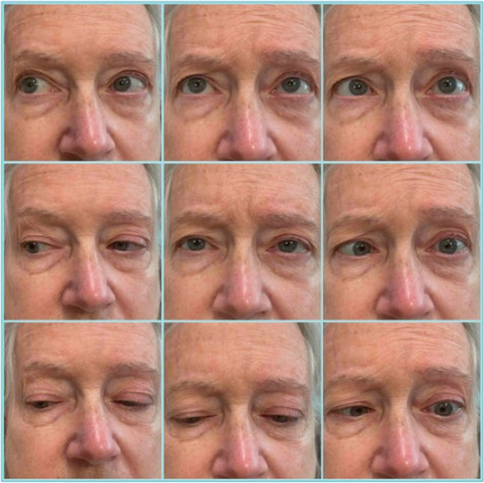
Case Presentation: A 73-year-old female with a history of hypertension and migraines presented to the ER with nausea, headache, and photophobia. She underwent CTA which showed a small right ICA aneurysm with no other acute abnormalities. She was treated and released but returned to the ER the next day with worsening symptoms. LP was unremarkable. UA revealed pyuria, so the patient was treated with ceftriaxone for 3 days. Her symptoms improved and she was discharged. A week later, she returned to the ER with double vision. MRI brain at the time was unremarkable. She was transferred to a tertiary hospital for further workup. Exam revealed left lid ptosis and left abducens palsy. WBC was 18.8 x 10⁹/L, CRP was 19.9 mg/dL and ESR was 51 mm/hr. MRI orbits revealed skull base osteomyelitis extending along the clivus with severe sphenoid and posterior ethmoid paranasal sinus disease. It also showed a 12 x 7 x 9 mm hypo-enhancing mass within the pituitary gland concerning for pituitary abscess. CT venogram head showed limited venous enhancement of the cavernous sinuses bilaterally, consistent with cavernous sinus thrombosis. Ophthalmology, ID, ENT, NSU, and endocrinology were consulted. ENT took the patient to the OR and identified excess pus in the sinuses. Blood cultures yielded viridans group streptococci and Staphylococcus epidermidis. Morning ACTH was 2 pg/mL, morning cortisol was 2.8 µg/dL, TSH was 0.61 µIU/mL, total T4 was 4.2 µg/dL, and total T3 was 22 ng/dL, concerning for central hypoadrenalism and hypothyroidism. She was diagnosed with skull base osteomyelitis and left cavernous sinus thrombosis causing a pituitary abscess. She was treated with IV ceftriaxone and vancomycin for six weeks and apixaban 5mg daily for cavernous sinus thrombosis. For central hypoadrenalism and hypothyroidism, she was treated with hydrocortisone and levothyroxine. Osteomyelitis and pituitary abscess resolved in follow-up imaging.
Discussion: This case illustrates two unusual conditions, pituitary abscess and Cavernous Sinus Thrombosis (CST), arising from a delayed headache diagnosis.A pituitary abscess is a rare, under-recognized cause of headaches. It can be classified as either primary or secondary in etiology.1 Primary pituitary abscesses develop from normal pituitary tissue while secondary pituitary abscesses develop from pre-existing pituitary lesions.2 The most common presenting symptoms are headache and visual defects. Pre-operative endocrinological abnormalities emphasize panhypopituitarism. Infectious symptoms are uncommon in most case reports of pituitary abscesses. CST occurs when the dural venous sinus becomes infected and thrombosed, producing a headache along with swelling around the eye and cranial nerve deficits.3 Infections of the face, such as the sphenoid and ethmoid sinusitis, can spread to the cavernous sinus. Early signs of CST are non-specific; however, cranial nerve signs on physical exam should prompt imaging studies. Staphylococcus aureus accounts for many cases associated with facial infection or sphenoid sinusitis.4 For hospitalists, early diagnoses and treatment are crucial to prevent morbidity and mortality.5
Conclusions: Headaches are common complaints in hospitalized patients; however, the presence of fever and ophthalmic findings warrants further investigation by hospitalists. In this case, the patient presented with a headache, ophthalmoplegia, and elevated inflammatory markers. CST and pituitary abscess should be considered in the differential diagnosis of headaches.