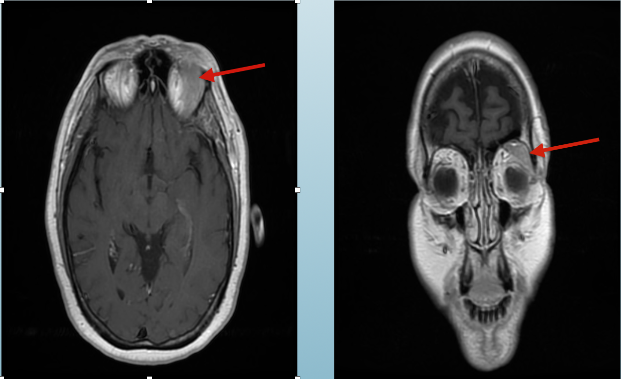
Case Presentation: A 62-year-old female with a past medical history of depression, obesity status postbariatric surgery, non-cardiac syncope and recurrent sinusitis presented to her primary care physicianfor further workup of her syncope. During this visit she was also complaining of continued left frontaland maxillary sinus pain for the past four weeks. She had recently completed a treatment course withamoxicillin/clavulanate for bacterial sinusitis with incomplete resolution of her symptoms. She endorsedpurulent nasal discharge, headache, malaise, left ear fullness, myalgia, fatigue and subjective fevers. Shefurther stated that she had suffered 3 episodes of sinusitis in the past 12 months. Examination revealedmild left periorbital edema with slight left proptosis, and left maxillary sinus pain. An MRI of the brainfor her syncope workup revealed a 3.1 x 2.3 x 1.3 cm lacrimal mass. Subsequent biopsy showed positiveB cell CD 19, 20, 22 and kappa light chain consistent with extranodal MALToma. PET scan showed noother tissue involvement, and bone marrow biopsy was normal signifying a stage 1 MALToma. Thepatient received localized radiation therapy and is currently undergoing post radiation surveillance
Discussion: Lacrimal gland MALTomas are a rare manifestation of primary extranodal Marginal B CellLymphoma, accounting for 1% of all Non-Hodgkin lymphoma subtypes. Malignant neoplasms of thelacrimal gland account for just 4% of all lacrimal gland lesions, 50% of which are adenoid cysticcarcinoma. The pathophysiology of MALTomas arising from the lacrimal gland remains unknown. Thesetumors are bimodal, typically presenting within the 3rd or 6th decade of life. They present withproptosis, vision changes, headache and periorbital numbness. Diagnosis is with biopsy showinglymphocytic proliferation. Treatment is based on staging however most are treated with directradiotherapy given they are often diagnosed at stage 1-2. The 5-year survival rate overall is 75%.
Conclusions: Literature on primary lacrimal gland tumors remains sparse due to the infrequency ofthese lesions. Primary lacrimal gland lymphomas are rare, and represent approximately 8-27% of ocular adnexa lymphomas. The mostcommon lymphoma affecting the lacrimal gland is a mucosa-associated lymphoid tissue lesion(MALToma). These lesions represent only 1% of all extranodal lymphomas, and the incidence of thesetumors is unknown. Additional studies on these lesions is needed to further understand these rare lesions.