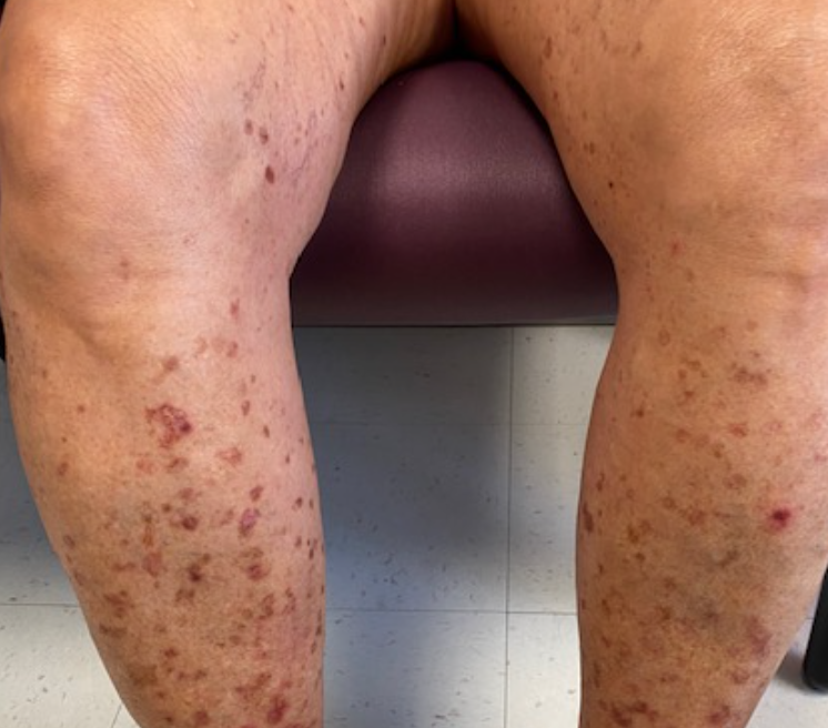
Case Presentation: A 60-year-old-female with past medical history of type 2 diabetes mellitus, hypertension, and hyperlipidemia presented with scaly pink macules with collarette ring of scale on bilateral lower extremity and forearms (figure 1). Some of her associated symptoms included itchiness. There was no drug use history and family history negative for similar skin lesion. She was recently started on gapabentin about one month ago and presumed diagnosis for skin lesion was erythema multiforme. The gabapentin was discontinued and patient was initially treated with 1% hydrocortisone cream and subsequently oral prednisone. However, patient’s skin lesions continued to worsen after 2 weeks of treatment. Patient was referred to dermatologist and patient had right lower extremity skin biopsy that showed superficial fragments of benign skin with focal parakeratosis. Negative for malignancy and no fungal organisms seen on periodic acid-schiff (PAS) stain. She was diagnosed with disseminated superficial actinic porokeratosis (DSAP) and started on 5-fluorouracil (5-FU) topical cream twice a day for 2 weeks. Photoprotective measures such as wearing long sleeves and sunscreen was encouraged.
Discussion: DSAP is a benign intraepidermal condition caused by sun exposure or genetic predisposition. While the incidence and prevalence of DSAP is unknown, an awareness of DSAP and its clinical features is important for appropriate diagnosis as skin related consultation account of for 12.4% of diseases seen by general practitioner (1). The lesions in DSAP start as pink to brown papules and macules with a raised border in sun exposed areas that can be asymptomatic or mildly pruritic (2). The sporadic form of DSAP is characterized by late onset and negative family history of the condition (3). In this particular case, sun exposure is the most likely primary etiologic factor for the development of porokeratotic lesions. The diagnosis of DSAP is usually clinically, with the help of dermatoscopic evaluation. DSAP have been associated with other disorders such as chronic liver disease, human immunodeficiency, and autoimmune diseases treated with systemic steroids (2). Also, there is a risk of malignancy developing from DSAP lesion. Malignant transformation occurs in 6.9% to 11.6% of patients and most common is transformation to squamous cell carcinoma. Treatment options include 5-fluorouracil, diclofenac, cryotherapy, imiquimod, photodynamic therapy,retinoids, vitamin D3 analogs. and topical lovastatin. Photoprotective measures should be recommended and patients need to follow up on regular basis for monitoring of their lesions (4).
Conclusions: Porokeratosis is a rare skin disorder that presents as keratotic papules or annular plaques with thin, raised border. It occurs on sun exposed skin and risk factors include genetics, immunosuppression, and ultraviolet light. Our case presentation looks at elderly patient who presents with skin rash and was diagnosed with DSAP. This case highlights the importance of recognizing DSAP and its clinical appearance given dermatologic conditions are frequent complaints. However, it is difficult to manage as treatment is poorly standardized and often not effective. In conclusion, it is necessary to further investigate the mechanism, risk factors, prevention and treatment of DSAP.