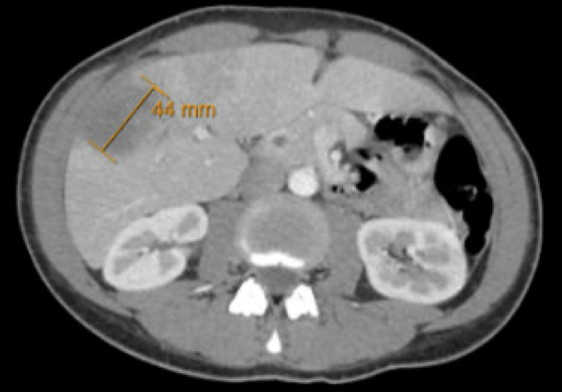
Case Presentation: A 32-year-old female patient without significant past medical history presented with one week of fevers to 102°F, right upper quadrant (RUQ) pain, and non-bloody diarrhea. She denied consuming unusual foods or traveling immediately before the onset of symptoms. However, she had traveled to Mexico, Denmark, South Korea, Guatemala, and Tanzania in the previous few years. She had been taking a combined oral contraceptive (OCP) for several years. She denied a personal or family history of blood clots, though her mother had an ischemic stroke in her 50s. On arrival, she was septic (febrile to 102.7°F, HR 94, WBC 12.6 K/µL) with mildly tender RUQ. She was found to have a heterogeneous hypoattenuating lesion of the right hepatic lobe (5.4 x 5.8 x 3.3 cm) and right hepatic vein thrombosis (HVT) on CT abdomen/pelvis and MRI abdomen. The GI pathogen panel was positive for Entamoeba histolytica, supporting the diagnosis of amoebic liver abscess. The patient was treated with oral metronidazole followed by paromomycin for intraluminal coverage. She was also started on a therapeutic heparin drip before conversion to apixaban for treatment of the hepatic vein thrombosis.
Discussion: Given the rarity of this presentation, we did not initially attribute the patient’s hepatic vein thrombosis to her liver abscess but rather placed more weight on OCP use versus possible thrombophilia. Her mother’s ischemic stroke at a relatively young age, paired with the fact that she had been on an OCP for years without incident (venous thromboembolism risk is notably highest in the first year of OCP use), led us down a diagnostic path of working up thrombophilia with selective hypercoagulable labs (Janus kinase 2 (JAK2) V617F mutation, Factor V Leiden (FVL) mutation, Dilute Russell viper venom time (DRVVT)). We specifically deferred ordering proteins C and S and antithrombin as they were acute-phase reactants. DRVVT was found to be weakly positive. We later learned that the weakly positive DRVVT was not interpretable given concurrent heparin use. Anticoagulants can directly prolong DRVVT, yielding significant rates of false positive results. Further, hepatic vein thrombosis is a rare but recognized complication of amoebic liver abscesses. In light of clear provoking factors, additional hypercoagulable workup was deemed unnecessary by hematology. Instead, emphasis was placed on the treatment of provoked HVT for at least three months until outpatient follow-up to assess for resolution.
Conclusions: With interesting presentations, there is sometimes an overzealousness in the pursuit of diagnostic clarity. In this case, ordering hypercoagulable studies was a well-intentioned course that could have carried long-term implications. If the weakly positive DRVVT portended the diagnosis of antiphospholipid syndrome, this young patient may have been subjected to indefinite warfarin therapy. Instead, we redirected our attention to this patient’s unique circumstances and tailored her treatment in a manner that was thoughtful and evidence-based. Therefore, considering the clinical context (HVT associated with abscess) and the applicability of respective studies (the interpretability of DRVVT in the setting of heparin use) may save both the patient and the medical team unnecessary emotional or financial burden. This case highlights that practicing diagnostic stewardship in a landscape of resource abundance may ultimately be the model of conscientious, high-quality care.