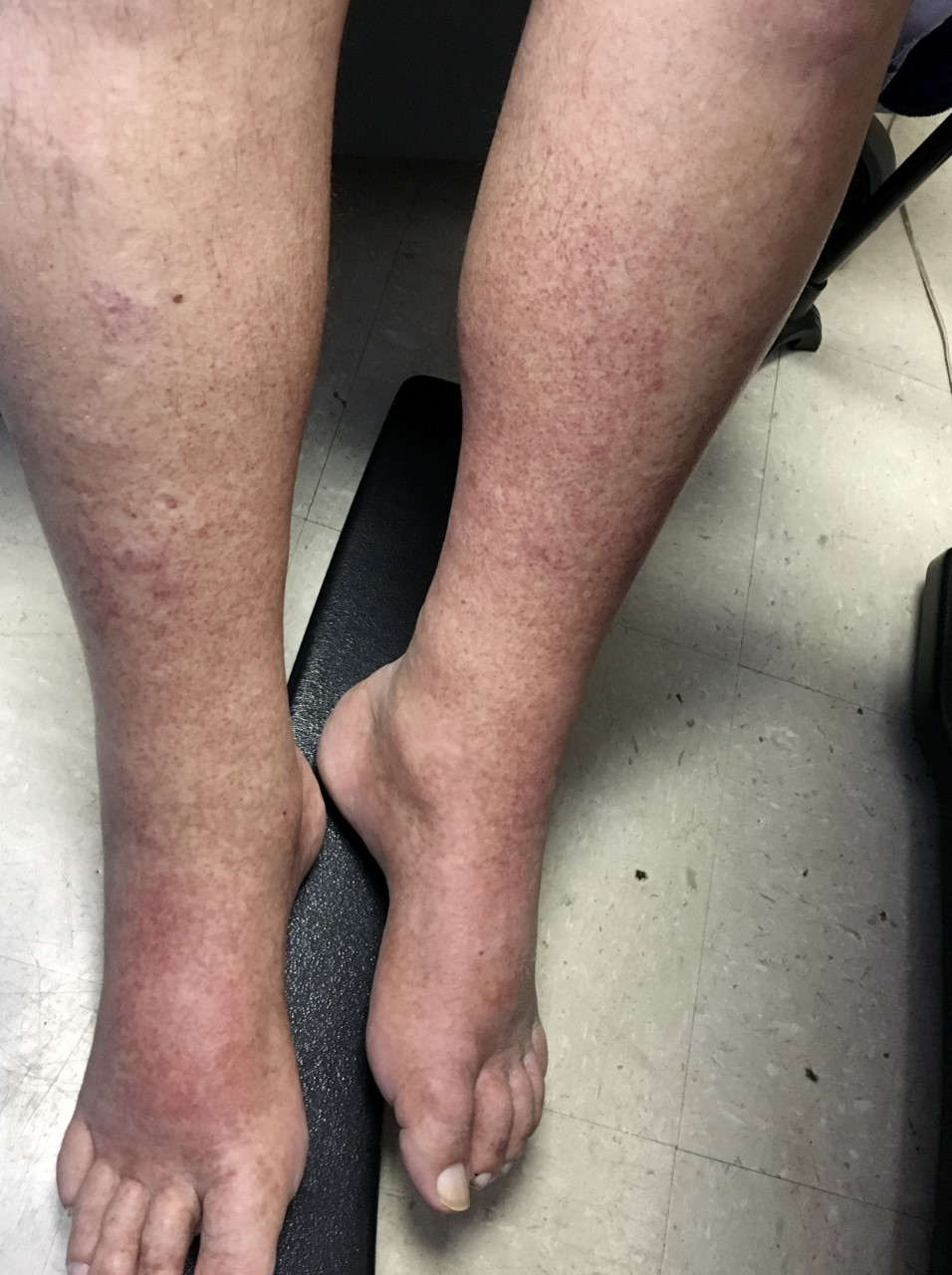
Case Presentation: 73 year old male with solitary kidney due to prior renal cell carcinoma and splenic marginal zone lymphoma currently treated with chlorambucil presented to the emergency department with acute kidney injury. He complained of red “kool-aid” urine and lower extremity rash. The rash was non-blanching, erythematous, papular and present on his ankles and extended circumferentially to his knees. He denied other symptoms including fever, arthralgia, neuropathy, or flank pain. On presentation, his creatinine was elevated to 7.5 (base-line 1.1), potassium was 6.0, and phosphorus was 7.7. Uric acid was 10, but decreased from prior. His LDH and total bilirubin levels were normal. His WBC was 4.5, Hgb 8.2, and platelets were 71, all stable. Urinalysis revealed large blood and nephrotic-range proteinuria. Renal ultrasound demonstrated mild hydronephrosis of his solitary kidney; computed tomography confirmed unilateral obstruction secondary to bulky lymphadenopathy. Urologic intervention improved but did not resolve renal dysfunction. Given purpura, proteinuria, and persistent kidney injury, further workup was pursued, which demonstrated low C3 and C4 levels, a positive rheumatoid factor and IgM predominance on SPEP. A skin biopsy revealed a resolving leukocytoclastic vasculitis. He was diagnosed with mixed cryoglobulinemia secondary to lymphoma and started on rituximab with resolution of kidney injury and rash.
Discussion: Acute kidney injury in the setting of a lymphoproliferative disorder increases mortality, delays cancer therapy, and provides a unique differential diagnosis. As in non-cancer AKI, the most common etiology is pre-renal etiology. Here, we discuss a framework describing the remainder of AKI in hematologic malignancy. Consider four categories of AKI related to the cancer itself: direct renal infiltration, obstructive uropathy from tumor burden or bulky lymphadenopathy, malignancy associated glomerular diseases, and malignancy-associated vasculitides. Treatment-induced acute kidney injury differentials should include: chemoradiotherapy toxicity (including tumor lysis syndrome) and acute tubular necrosis from sepsis. Imaging with ultrasound or CT may be helpful; however, history, physical exam, and urine sediment evaluation are crucial.This case highlights malignancy-associated vasculitis as an etiology to consider in AKI in a patient with hematologic malignancy, particularly with purpura. Diagnosis of mixed cryoglobulinemia is based on the presence of cryoglobulins in the blood coupled with the clinical presentation. Mixed cryoglobulinemias are most notoriously associated with HCV infection (~80%) but are also associated with lymphoproliferative disorders, autoimmune disease, and other infections. Purpura is the most suggestive feature (90% of cases) but arthralgias, peripheral neuropathy, and glomerulonephritis are also suggestive. Supportive laboratory features include hypocomplementemia (specifically C4), a positive rheumatoid factor, and hypergammaglobulinemia.
Conclusions: Acute kidney injury in patients with hematologic malignancy presents a targeted differential diagnosis for the hospitalist, which includes kidney injury related to the cancer itself or the unique treatments thereof. Cryoglobulinemic vasculitis should be considered in patients with underlying leukemia/lymphoma presenting with renal dysfunction and purpura.