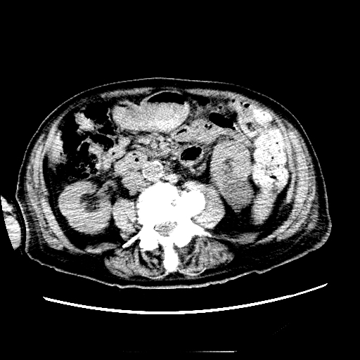
Case Presentation: An 87 year old male with a history HFrEF 35%, BPH, Afib, seizure disorder. He had a recent fall and presented with A-fib with RVR. His creatinine at that time was found to be 5.6 (no baseline creatinine at that time). Renal and bladder ultrasound showed large intravesical mass consistent with bladder mass versus prostate mass versus clot. Foley catheter was placed and kidney function improved after bladder irrigation. Pt was discharge to NH. Creatinine at time of discharge was 2.9. In nursing home he had vomiting and was found to be very lethargic, having metallic taste in mouth and decrease in urine output. Pt denied fever, chills, cough, SOB. Blood work showed Creatinine of 6.5 with metabolic acidosis. Urine sediment showed few coarse granular casts. Pt underwent one session of dialysis. Lab work up showed lymphocytosis, anemia, and free light chains were positive. Imaging studies showed splenomegaly and retroperitoneal lymphadenopathy. Given the unclear nature of his AKI he underwent kidney biopsy that showed infiltration of the kidney with lymphocytes and chronic parenchymal changes with 46% global Glomerulosclerosis CD20 and CD5 positive and flow cytometry of the peripheral blood reveals CLL, suggesting that lymphoma is the likely etiology of his renal failure. He was started on steroids and showed improvement of his renal function.
Discussion: Chronic lymphocytic leukemia (CLL) is characterized by a lymphocytosis of mature-appearing clonal CD5+, CD23+ B lymphocytes. CLL cells arise from the bone marrow and infiltrate lymphoid tissues such as lymp nodes and spleen. Presentation is usually through discovery of lymphocytosis or lymphadenopathy. Unusual presentations, especially paraneoplastic syndromes are rare. Here, we describe a case of severe acute kidney injury (AKI) associated with the presence of a monoclonal protein in serum. Workup for suspected plasma cell dyscrasia led instead to the diagnosis of bone marrow infiltration by atypical CLL with lymphocytosis. Renal biopsy showed a glomerulonephritis turned out to be paraneoplastic as it went into remission after treatment for CLL in the context of seemingly unrelated conditions that may be paraneoplastic in origin.
Conclusions: Our case shows an unusual presentation of CLL and prompts for increased awareness of lymphoproliferative disorders. The case report strongly underlines the utility of performing a bone marrow biopsy in patients presenting an abnormal electrophoresis, immunofixation or free light chain ratio, even in the absence of any signs of CLL from physical examination and blood analysis to detect a lymphoproliferative disorder. Because of our patient’s age and renal impairment, we started treatment with a steroid pretreatment phase which are known to be efficacious in CLL. Our patient developed good response on steroid therapy.
.jpg)