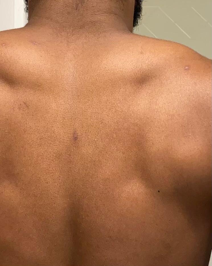
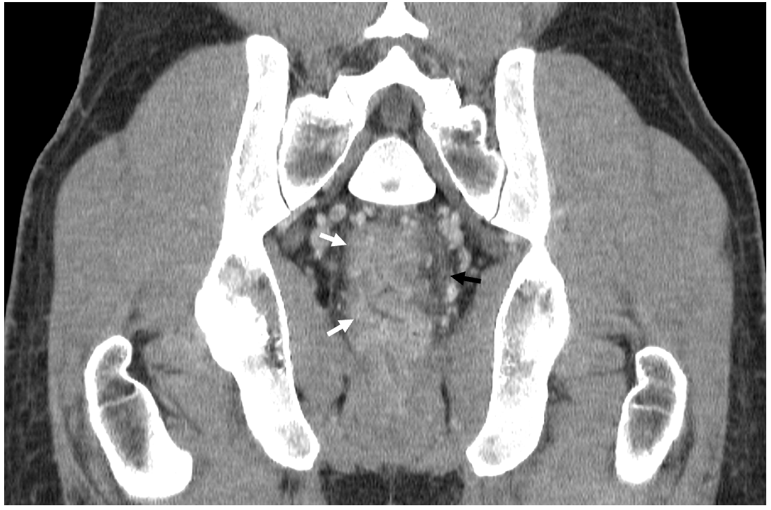
Case Presentation: A 33-year-old man with a history of human immunodeficiency virus (HIV, 2007, well-controlled on antiretrovirals) and recent rectal chlamydia (on routine screening) presented to the emergency department (ED) with a five-day history of profuse rectal bleeding, pain, and tenesmus; preceded by a prodrome of fever, myalgias, productive cough, and a painful papular skin rash. The papules were not grouped, appeared at different timepoints, were diffuse (head, mouth, trunk, genitals, palms and soles), and underwent asynchronous crusting. Sexual practices included receptive anal intercourse with multiple partners without the use of barrier protection, the most recent of which was three weeks prior to presentation. No recent travel history. Upon examination, he was afebrile with diffuse papular and pustular skin lesions on the trunk and extremities, as well as the scrotum and base of the penis; no perianal lesions. Hemoglobin was 12.5 mg/dL. Initial pelvic computed tomography (CT) demonstrated inflammatory proctitis with bulky adenopathy. Skin lesions swab eventually returned positive for monkeypox virus polymerase chain reaction (PCR). He received treatment for chlamydia and antiviral therapy (tecovirimat), with subsequent improvement in rectal bleeding and rash. He was discharged to self-isolation at home.
Discussion: The current monkeypox pandemic involves a high proportion of men-who-have-sex-with-men (MSM), suggesting possible inoculation via the anogenital region, as rectal abrasions might lead to a deeper inflammatory process. Our patient exhibited rectal wall inflammation on CT imaging, which may have progressed to profuse bleeding. Additionally, coexistent “dormant” sexually transmitted infections (STI) might have primed and led to friable rectal wall tissue leading to bleeding. One cohort suggests monkeypox has coexisting STIs in 76%, so STI screening remains paramount. Maintaining a low threshold for suspecting monkeypox in the appropriate clinical setting and in at-risk populations is important for timely intervention.
Conclusions: The current monkeypox outbreak has seen maculopapular lesions at ports of entry (e.g. skin, mucosal surfaces) followed by more invasive and severe inflammatory manifestations—including in the pharynx and the rectum—in some patients, including proctitis in up to 25% in a recent cohort. Indications for tecovirimat (TPOXX®) therapy include patients with severe disease (e.g. hemorrhagic disease, confluent lesions, sepsis, encephalitis, or requiring hospitalization) or infections involving lesions in mucocutaneous sites which may constitute a special hazard (e.g. eyes, mouth, genitals, or anus). Our patient with monkeypox and proctitis, rapidly received pharmacologic therapy. Curbing transmission—through education, pre-exposure vaccination, timely identification, and isolation—in communities at risk, while avoiding stigmatization, are necessary to control this emerging public health outbreak.


